
The fastest way to cure blepharitis is to treat its root cause – dry eyes, blepharitis, meibomian gland dysfunction all at the same time. The goal is to get the inflammation under control. TheraLife designed a protocol for blepharitis relief that is comprehensive and prevents it from recurring.
My personal story of Blepharitis MGD Recovery
“I have had blepharitis that would come concurrent with a pink eye for quite a few years, but it has always gone away pretty quickly on its own. However, this last time, blepharitis and dry eye did not leave, which became quite disruptive to my life. My eye doctor gave me eye drops, which only made my eyes feel drier. I felt as though I had no hope for my eyes, and they were going to keep getting worse and worse. Despite being fairly skeptical, I finally decided to try Theralife, and I’m So glad I did. I’ve now been taking it for just over two weeks, and I already feel so much better. This morning I woke up, and my eyes were almost white instead of bright red. My blurry vision cleared up, the stickiness was gone from my eye, and the stinging had almost left. I’m looking forward to them getting better and better. Thanks so much!”
Cindy, USA
The TheraLIfe Solution to a Blepharitis Cure

What is blepharitis
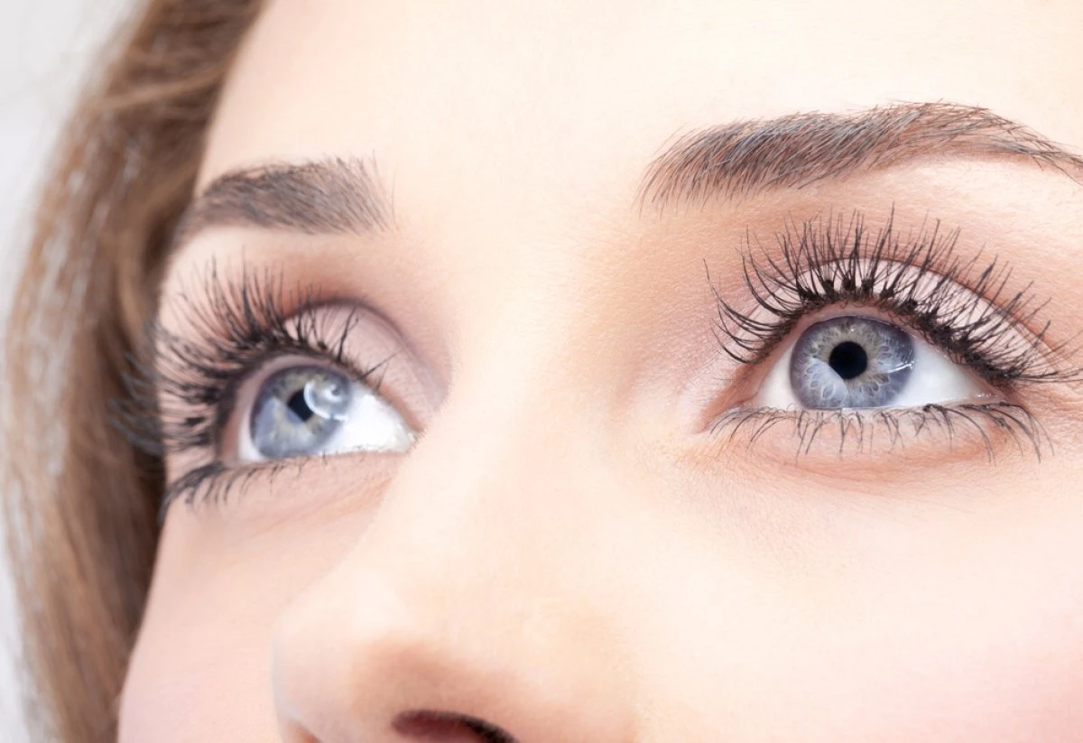
Blepharitis is the inflammation of the eyelids. Blepharitis usually affects both eyes along the edges of eyelids.
Blepharitis refers to a family of inflammatory disease processes of the eyelid(s).
Types
Blepharitis can be divided anatomically into anterior and posterior blepharitis.
- Anterior blepharitis refers to inflammation mainly centered around the skin, eyelashes, and lash follicles. Anterior blepharitis has staphylococcal and seborrheic variants.
- Posterior blepharitis involves the meibomian gland orifices, meibomian glands, tarsal plate, and blepharo-conjunctival junction.
Causes
- Chronic dry eyes
- Acne rosacea. Rosacea causes facial skin inflammation, including eyelids.
- Allergies. Allergies to contact lens solution, eye drops, or makeup can spur irritation.
- Dandruff (Seborrheic dermatitis). …
- Dry eye.
- Lice or mites in eyelashes (Demodicosis).
Blepharitis is often a chronic condition that’s difficult to treat. Blepharitis can be uncomfortable and unsightly. But it usually doesn’t cause permanent damage to your eyesight, and it’s not contagious.
Poor hygiene
Blepharitis is a chronic or long-term inflammation of the eyelids and eyelash hair follicles. It affects people of all ages. The most common causes of blepharitis are:
- poor eyelid hygiene;
- excessive oil produced by the glands in the eyelid;
- bacterial infection (often staphylococcal)
- allergic reaction.
Blepharitis is associated with chronic diseases.
People With Blepharitis are more Likely to have certain Inflammatory Diseases, Psychological Issues, Cardiovascular Diseases and More.
Getting rid of blepharitis is to control the inflammation, which will keep these diseases in check.
What chronic diseases
Several eye and systemic problems are more common among people with chronic blepharitis, but the reasons aren’t always clear.
Associated with blepharitis was the presence of :
- Inflammatory diseases (such as gastritis and asthma),
- Psychological problems (such as anxiety and depression),
- Hypothyroidism
- Cardiovascular diseases
- Chalazia/chalazion
- Pterygium – noncancerous growth in the conjunctiva
- Ulcerative colitis
- Irritable Bowel Syndrome
- Rosacea
- Seborrheic dermatitis
- Trichasis – miss directed eyelashes
- Ectropion and entropion- Lower eyelid turn outward or inward.
- Conjunctivitis
- Keratitis – inflammation of the cornea
- Autoimmune diseases- Sjogren’s, Lupus, Rheumatoid Arthritis.
Pathology
Blepharitis frequently involves bacterial colonization of the eyelids.
Seborrheic dermatitis and meibomian gland dysfunction have increased colonization of the lid margin. Which results in the direct microbial invasion of tissues, immune system-mediated damage or damage caused by the production of bacterial toxins, waste products, and enzymes.
Statistics
Frequency
United States
Blepharitis is a common eye disorder in the United States and throughout the world. 86% of all patients with dry eyes also have blepharitis. More than 25 million Americans suffer from blepharitis.
Mortality/Morbidity
The exact association between blepharitis and mortality is not known. The disease process can result in damage to the lids with trichiasis, notching entropion, and ectropion. Corneal damage can result in inflammation, scarring, loss of surface smoothness, irregular astigmatism, and loss of optical clarity. If severe inflammation develops, corneal perforation can occur.
Race
No known studies demonstrate racial differences in the incidence of blepharitis. Rosacea may be more common in fair-skinned individuals.
Sex
No differences in blepharitis incidence and clinical features between the sexes have been found.
Age
Seborrheic blepharitis is more common in an older age group. The apparent mean age is 50 years.
Prognosis
Overall, the prognosis for patients with blepharitis is good to excellent.
Blepharitis only causes significant morbidity in a tiny subset of patients. For most, it remains more of an asymptomatic affliction than a genuine threat to their health and function.
Impact of chronic blepharitis on quality of life.
People with chronic blepharitis experience discomfort and misery that can significantly reduce their well-being and ability to carry out daily life and work activities. Recognition of the off and on course of the disease, and the necessity of management through a prolonged program rather than an instant cure, help them successfully approach the condition.
Clinical presentation.
Blepharitis typically presents with eye irritation, itching, red eyelids, flaking of the lid margins, or changes in the eyelashes.
Other common complaints include the following:
- Burning
- Watering
- Foreign body sensation
- Crusting and mattering of the lashes and medial canthus
- Red lids
- Red eyes
- Photophobia – light sensitivity
- Pain
- Decreased vision
- Visual fluctuations
- Heat, cold, alcohol, and spicy-food intolerance
The condition most typically has a chronic course with intermittent exacerbations and eruptions of symptomatic disease.
Physical Findings
External examination of people with blepharitis often demonstrates findings of associated conditions.
Herpes skin disease can be associated with red and blister formation.
Seborrheic dermatitis
Seborrheic dermatitis is typified by oily skin and flaking from the scalp or brows.
Rosacea
Rosacea is associated with pimples, a red nose, spider veins of the cheeks and eyelid margins, erythema, and papules.
Clinical features
Gross examination of the eyelids shows erythema and crusting of the lashes and lid margins.
Slit-lamp examination shows additional features:
- Loss of lashes (madarosis),
- Whitening of the lashes (poliosis),
- Lid scarring and misdirection of lashes (trichiasis),
- Crusting of the lashes and meibomian orifices,
- Eyelid margin ulceration,
- Plugging and “pouting” of the meibomian orifices,
- Telangiectasias of the lid margin,
- Lid irregularity (tylosis).
- The conjunctiva usually shows papillary injection. Advanced cases reveal tarsal thickening,
- Loss of normal tarsal vascular architecture,
- Subconjunctival substantia propria fibrosis,
- Conjunctival scarring
- Tarsal distortion due to cicatricial contraction and subsequent entropion.
Corneal findings can include:
- Ounctate epithelial erosions,
- Marginal infiltrates,
- Marginal ulcers,
- Limbal inflammation and thickening (limbitis),
- Peripheral corneal ectasia, pannus, and phlyctenule formation.
Corneal can progress to infection and even perforation.
Anterior Blepharitis associated with bacterial infections.
The anterior variant of blepharitis involves mainly the lashes and associated non-meibomian oil glands. Various formations of debris adhere to the lashes.
- Crusting refers to flakes of material that adhere to the lashes and usually represent seborrheic disease. The epithelial material is the crusting
- In staphylococcal diseases, a collarette is an irregular ringlike formation. Staphylococcal blepharitis has the formation of collarettes on the lashes.
- A sleeve is a smooth tube of material that also surrounds the base of the lash as it intersects the lid. Sleeving is associated with infection by the eyelash parasite,
- Ulcers at the base of the lashes.-covered by a crust of fibrin, which is lifted as the lash shaft grows.
- Seborrheic blepharitis also involves the anterior lid primarily. It is associated with the formation of greasy crusts of material adherent to the eyelash shaft.
Corneal disease is most familiar with the staphylococcal variant of anterior lid disease.
Posterial Blepharitis associated with MGD
Posterior blepharitis is principally related to dysfunction of the meibomian glands. Alterations in secretory metabolism and function lead to disease. The meibomian secretions become more waxlike and begin to block the gland orifices. The stagnant material becomes a growth medium for bacteria and can seep into the deeper eyelid tissue layers, causing inflammation. The inflammation leads to gland plugging, hardening of the lipid secretory material, inflamed orifices, and the formation of chalazia.
Complications
Conjunctivitis and keratitis(inflammation of the cornea) are common. It can result in a complication of blepharitis and require additional treatment besides eyelid margin therapy.
Antibiotics
Antibiotic-corticosteroid solutions can significantly reduce inflammation and symptoms of conjunctivitis. Corneal infiltrates are with antibiotic-corticosteroid drops.
Small marginal ulcers are treated empirically. Larger, paracentral, or atypical ulcers should be scraped, and specimens sent for diagnostic slides and culture and sensitivity testing.
The use of steroids can lead to cataracts, glaucoma, and viral reactivations.
Recurrent bouts of inflammation and scarring from blepharitis can promote positional eyelid disease. Trichiasis and lid notching can result in keratitis and severe symptoms. These conditions often are very refractory to simple management steps.
Entropion or ectropion can develop and complicate the clinical situation.
Laboratory Tests
In general, there is no need for diagnostic tests for suspected blepharitis. Research and other rare protocols may involve:
- Eyelid margin cultures.
- Transillumination studies of the meibomian glands.
- Digital-imaging techniques.
- Conjunctival impression cytology.
- Marginal biopsies.
- Even analysis of gland secretions.
We recommend testing people with blepharitis for tear insufficiency or nasolacrimal drainage problems.
To learn more, call toll free 1-877-917-1989 US/Canada
send email to [email protected]
References
- Korb, D. R. & Henriquez, A. S. Meibomian gland dysfunction and contact lens intolerance. J. Am. Optom. Assoc.51, 243–251 (1980).
2.Nelson, J. D. et al. The international workshop on meibomian gland dysfunction: report of the definition and classification subcommittee. Invest. Ophthalmol. Vis. Sci. 52, 1930–1937.
3.Eom, Y., Lee, J. S., Kang, S. Y., Kim, H. M. & Song, J. S. Correlation between quantitative measurements of tear film lipid layer thickness and meibomian gland loss in patients with obstructive meibomian gland dysfunction and normal controls. Am. J. Ophthalmol. 155, 1104-1110 e1102. 4.
- Bron, A. J. et al.TFOS DEWS II pathophysiology report. Ocul. Surf.15, 438–510.
- Craig, J. P. et al.TFOS DEWS II definition and classification report. Ocul. Surf.15, 276–283.
- Mathers, W. D. Ocular evaporation in meibomian gland dysfunction and dry eye. Ophthalmology100, 347–351 (1993).
- Georgiev, G. A., Eftimov, P. & Yokoi, N. Structure-function relationship of tear film lipid layer: a contemporary perspective. Exp. Eye Res.163, 17–28. 8.
- Sledge, S. M. et al.Evaporation and hydrocarbon chain conformation of surface lipid films. Ocul. Surf.14, 447–459.
- Han, K. E. et al.Evaluation of dry eye and meibomian gland dysfunction after cataract surgery. Am. J. Ophthalmol.157, 1144-1150 e1141.
- Jung, J. W. et al.Meibomian gland dysfunction and tear cytokines after cataract surgery according to preoperative meibomian gland status. Clin. Exp. Ophthalmol.44, 555–562. 11.
- Lee, J. A. & Cho, Y. K. The influence of preoperative meibomian gland disease on dryness after cataract surgery. J. Korean Ophthalmol. Soc.57, 228–235 (2016).
- Park, Y., Hwang, H. B. & Kim, H. S. Observation of influence of cataract surgery on the ocular surface. PLoS ONE11, e0152460. 13.
- Starr, C. E. et al.An algorithm for the preoperative diagnosis and treatment of ocular surface disorders. J. Cataract. Refract. Surg.45, 669–684. 14.
- Yoo, S. E., Lee, D. C. & Chang, M. H. The effect of low-dose doxycycline therapy in chronic meibomian gland dysfunction. Korean J. Ophthalmol.: KJO19, 258–263. 15.
- Nam, S. W., Lim, D. H., Hyun, J. & Chung, T.-Y. Effects and prognostic factors of automated thermodynamic system treatment for meibomian gland dysfunction. J. Korean Ophthalmol. Soc.57, 724–733 (2016).
- Key, J. E. A comparative study of eyelid cleaning regimens in chronic blepharitis. CLAO J.22, 209–212 (1996).
- Paranjpe, D. R. & Foulks, G. N. Therapy for meibomian gland disease. Ophthalmol. Clin. N. Am.16, 37–42 (2003).
- Borchman, D. The optimum temperature for the heat therapy for meibomian gland dysfunction. Ocul. Surf.17, 360–364.
19. Lindsley, K., Matsumura, S., Hatef, E. & Akpek, E. K. Interventions for chronic blepharitis. Cochrane Database Syst. Rev.
20. Benitez-Del-Castillo, J. M. How to promote and preserve eyelid health. Clin. Ophthalmol.6, 1689–1698.
- Guillon, M., Maissa, C. & Wong, S. Symptomatic relief associated with eyelid hygiene in anterior blepharitis and MGD. Eye Contact Lens38, 306–312. 22.
- Dougherty, J. M. & McCulley, J. P. Bacterial lipases and chronic blepharitis. Invest. Ophthalmol. Vis. Sci.27, 486–491 (1986).
- Peral, A., Alonso, J., Garcia-Garcia, C., Nino-Rueda, C. & Calvo Del Bosque, P. Importance of lid hygiene before ocular surgery: qualitative and quantitative analysis of eyelid and conjunctiva microbiota. Eye Contact Lens42, 366–370. 24.
- Hamada, S. et al.Assessment of the effect of cyclosporine-A 0.05% emulsion on the ocular surface and corneal sensation following cataract surgery. Contact Lens Anterior Eye39, 15–19. 25.
- Oh, T., Jung, Y., Chang, D., Kim, J. & Kim, H. Changes in the tear film and ocular surface after cataract surgery. Jpn. J. Ophthalmol.56, 113–118. 26.
- Kim, J. H., Chung, J. L., Kang, S. Y., Kim, S. W. & Seo, K. Y. Change in corneal sensitivity and corneal nerve after cataract surgery. Cornea28, S20–S25. 27.
- Nosch, D. S., Pult, H., Albon, J., Purslow, C. & Murphy, P. J. Relationship between corneal sensation, blinking, and tear film quality. Optom. Vis. Sci.93, 471–481.
- Wan, T., Jin, X., Lin, L., Xu, Y. & Zhao, Y. Incomplete blinking may attribute to the development of meibomian gland dysfunction. Curr. Eye Res.41, 179–185.
- Tsubota, K. Tear dynamics and dry eye. Prog. Retin. Eye Res.17, 565–596. 30.
- Knop, E., Knop, N., Millar, T., Obata, H. & Sullivan, D. A. The international workshop on meibomian gland dysfunction: report of the subcommittee on anatomy, physiology, and pathophysiology of the meibomian gland. Invest. Ophthalmol. Vis. Sci.52, 1938–1978.
- Eom, Y. et al.Comparison of meibomian gland loss and expressed meibum grade between the upper and lower eyelids in patients with obstructive meibomian gland dysfunction. Cornea33, 448–452.
41.Crosby, N. J., Shepherd, D. & Murray, A. Mechanical testing of lid speculae and relationship to postoperative ptosis. Eye (London) 27, 1098–1101.
- Marques-Fernandez, V. et al.An objective evaluation of the upper eyelid position after phacoemulsification cataract surgery. Semin. Ophthalmol.34, 442–445.
- Choi, Y. J. et al.Perioperative ocular parameters associated with persistent dry eye symptoms after cataract surgery. Cornea37, 734–739.
- Speaker, M. G., Milch, F. A., Shah, M. K., Eisner, W. & Kreiswirth, B. N. Role of external bacterial flora in the pathogenesis of acute postoperative endophthalmitis. Ophthalmology98, 639–649.
- Eom, Y. et al.Distribution and characteristics of meibomian gland dysfunction subtypes: a multicenter study in South Korea. Korean J. Ophthalmol.: KJO33, 205–213.
46. Viso, E., Rodriguez-Ares, M. T., Abelenda, D., Oubina, B. & Gude, F. Prevalence of asymptomatic and symptomatic meibomian gland dysfunction in the general population of Spain. Invest. Ophthalmol. Vis. Sci.53, 2601–2606.
- Bron, A. J., Evans, V. E. & Smith, J. A. Grading of corneal and conjunctival staining in the context of other dry eye tests. Cornea22, 640–650.
- Y., Lee, J. S., Keun Lee, H., Myung Kim, H. & Suk Song, J. Comparison of conjunctival staining between lissamine green and yellow filtered fluorescein sodium. Can. J. Ophthalmol.50, 273–277.
- Yuchs, J. Efficacy of topical azithromycin ophthalmic solution 1% in the treatment of posterior blepharitis. Adv. Ther.25, 858–870.
50.Efron, N. Efron Grading Scales for Contact Lens Complications (Millennium Edition). Butterworth-Heinemann (2000).
51.Arita, R. et al. Development of definitive and reliable grading scales for meibomian gland dysfunction. Am. J. Ophthalmol. 169, 125–137.
- Tomlinson, A. et al.The international workshop on meibomian gland dysfunction: report of the diagnosis subcommittee. Invest. Ophthalmol. Vis. Sci.52, 2006–2049.
53.Foulks, G. N. & Bron, A. J. Meibomian gland dysfunction: a clinical scheme for description, diagnosis, classification, and grading. Ocul. Surf. 1, 107–126 (2003).
54.Adil, M. Y. et al. Meibomian gland morphology is a sensitive early indicator of meibomian gland dysfunction. Am. J. Ophthalmol. 200, 16–25.
- Faul, F., Erdfelder, E., Lang, A. G. & Buchner, A. G*Power 3: a flexible statistical power analysis program for the social, behavioral, and biomedical sciences. Behav. Res. Methods39, 175–191.