Meibomian Gland Dysfunction (MGD)

Stop Recurring MGD Now With All-In-One-Starter Kit
- TheraLife Eye capsules to improve oil production in Meibomian Glands.
- Relief dry eyes to stop MGD/Blepharitis
- Stop recurring styes and chalazion
- Restore from the inside out, no more artificial tears.
Testimonial
“I was diagnosed with chronic dry eyes, blepharitis and meibomian gland dysfunction. My eyes were red, dry and painful. I tried many eye drops including prescription drops, antibiotic and steriod drops with no results. I started TheraLife Eye and can feel the difference in 2-3 weeks. I still cannot believe I have such total relief. I am so grateful for TheraLife.”
-C.L, Ohio
*Results may vary
Contains everything you need to get started in treating dry eye syndrome, blepharitis, and/or meibomian gland dysfunction (MGD).
All-in-One Starter Kit includes:
4 bottles of TheraLife® Eye Enhanced
1 bottle of Omega-3 Fish Oil
1 bottle Eye Lid Cleanser
1 Hot Compress
TheraLife® especially recommends this starter kit to Blepharitis/MGD dry eye sufferers. It includes all of the essential elements for getting lasting MGD/dry eye relief, collected for you into one conveniently packaged solution.
TheraLife® Eye Enhanced – clinically proven to restore normal tear production for those suffering from blocked meibomian glands, clogged meibomian glands, meibomian cysts (chalazion), meibomian blepharitis, meibomianitis, and other problems associated with meibomian gland disease. TheraLife® Eye Enhanced heals eye oil glands intracellularly – from the inside – in combination with our omega three supplements, warm compress, and eyelid cleanser.
TheraLife® Eye Enhanced has been clinically proven to restore normal tear production for those suffering from blocked meibomian glands, clogged meibomian glands, meibomian cysts (chalazion), meibomian blepharitis, meibomianitis, and other problems associated with meibomian gland disease.
TheraLife® Eye Enhanced heals eye oil glands intracellularly – from inside – in combination with our omega 3 supplements, hot compress, and eye lid cleansings. Buy the complete treatment dry eye relief kit today!
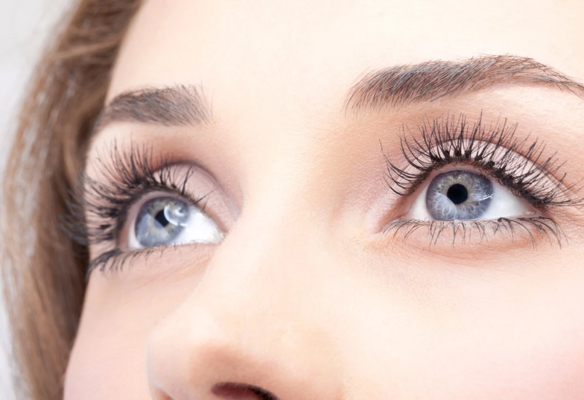
What is Meibomian Gland Dysfunction (MGD)?
Meibomian glands are located on the upper and lower eyelids, inside the lash line. Meibomian gland dysfunction (MGD) occurs when the oil glands in the eye become inflamed and blocked or clogged—resulting in reduced oil production. MGD causes posterior blepharitis.
Often concurred together with meibomian gland dysfunction are:
Chalazion
A chalazion (plural = chalazia) is like a bubble of fluid that forms under the eyelid if the eye’s normal secretions cannot be dispersed and become trapped in one pocket of tissue.
MGD and chalazion often occur together for most patients, combined with other problems, like blepharitis and dry, watery, crusty eyes.
Dry Eye Disease
Dry eye disease is the most common cause of MGD. The vision can dry due to a lack of proper lubrication, and tears evaporate too quickly. If the eye’s oil glands become clogged, a crust or other build-up will form.
Watery eyes
The eye can also become overly watery if it is trying to compensate for being too dry.
The problem is that if you have MGD, there are no oils to thicken the tears, meaning that the ones you do produce – in overabundance – will be of poor quality, too thin, and insubstantial to offer lasting natural relief.
Meibomian Gland Dysfunction (MGD) is very difficult to treat and is one of the most challenging eyes doctor issues.
How Do Tears Work?
When you blink, a film of tears spreads over the eye – keeps the eye’s surface smooth and clear.
The tear film has three layers:
- An oil layer
- A watery layer
- A mucus layer
Each layer serves a purpose.
The oil layer is the outside of the tear film. This layer is made in the eye’s meibomian glands. It makes the tear surface smooth and keeps the eyes moist. You get dry eye syndrome and evaporative dry eye when you produce less oil.
The watery layer is in the middle. It makes up most of what we see as tears. This layer cleans the eye, washing away particles that do not belong in the eye. This layer comes from the lacrimal glands in the eyelids.
Mucus is made in the conjunctiva – which is the clear tissue covering the white of your eye and inside your eyelids. The mucus layer is the tear film’s inner layer, which helps spread the watery layer over the eye’s surface, keeping it moist. Without mucus, tears would not stick to the eye.
Typically, our eyes constantly make tears stay moist. If our eyes are irritated, or we cry, our eyes make a lot of tears. But, sometimes the eyes don’t make enough, or something affects one or more layers of the tear film. In those cases, we end up with dry eyes.
Symptoms
The symptoms include burning that is worst on waking (by contrast, if you have aqueous deficient eyes, burning worsens as the day goes on). Increased tear evaporation leads to dry eyes, and MGD and dry eye symptoms go hand in hand. Treating the MGD can be the key to alleviating symptoms, such as:
- Grittiness
- Foreign body sensation
- Burning
- Dryness
- Light sensitivity
- Blurred vision
- Contact lens intolerance
Dry eye symptoms and MGD are always linked.
Diagnosis
The clinical presentation of MGD includes capped meibomian glands and foamy tears at the front surface.
Your eye doctor can perform meibomian gland expression to determine the extent of the damage to render treatment accordingly.
The secretions can be thickened and have a toothpaste appearance, making meibomian gland expression difficult. Ask your eye doctor what your secretions look like. Typically the eye doctor squeeze the lower lid.
The plugged glands can become infected, and the patient may complain of recurrent styes or chalazion.
The dysfunctional lipid layer in MGD leads to a rapid tear film break up and evaporative dry eyes.
Traditional Treatment Options
Always seek professional medical advice from your eye doctors before treatment.
The treatment options often involve:
No eye makeup
Avoid eye makeup to keep eyelid margins clean.
Warm compresses:
- To melt the clogged meibomian glands. Warm compresses help the abnormal meibum change from solid to liquid at a higher melting point than the surface temperature; when the meibum liquifies, the glands are no longer obstructed.
- Note a warm wet washcloth does not work. A warm washcloth does not get hot enough.
Lid scrubs and massage of eyelids –
- Clean your lid margin to prevent eyelid inflammation. Avenova works best because it disrupts the biofilm layer of the eyelids to prevent bacteria re-attachment, thus reducing inflammation.
- Gently massage both the upper lid and lower lid right after warm compresses.
- Note – baby shampoo does not work.
Omega 3 fish oil
To improve the poor quality of tear film in dry eye disease. Compensating for meibomian glands producing less oil.
Antibiotics
- Oral doxycycline (oral pill) has strong anti-inflammatory properties and softens oil glands’ plugs.
Steroid eye drops
- – for a duration of no more than one month. Long-term use cause glaucoma.
Physical probes– meibomian gland expression
Your eye doctor applies a topical anesthetic and uses cotton tip applicators, or physical instrument to apply pressure on your eyelids to perform meibomian gland expression to unclog your oil glands.
More advanced MGD treatment:
Many patients seek advanced treatment that works for them.
Laser treatment
- – Intense pulsed light for your eyes loosens clogging.
Thermal Pulsation Heat Wave Treatment–
- A series of 4 treatments that use heat to perform meibomian gland expression. Laser light is beneficial for people with rosacea.
Intraductal meibomian probing treatment
The most advanced mgd treatment. Very few eye doctors are trained to perform this procedure.
These MGD dry eye treatments are typically repeated every 6-9 months to maintain meibomian gland health.
Prognosis
According to the American Association of Opthalmology, in about 80% of cases, daily application of warm compresses to the eyelids followed by eyelid massage can help restore the normal consistency of the meibum, which in turn restores meibomian gland function keeping your eyes healthy.
MGD left untreated can cause severe damage to your cornea.
Note: Frequent meibomian gland expression at home is not recommended. It takes time for meibomian glands to regenerate.
Risk factors
Hormonal aspects
Androgen and estrogen receptors are present within meibomian glands, and meibocytes contain the enzymes necessary for the intracrine synthesis and metabolism of sex steroids.
Systemic Medications
Administration of Accutane is associated with severe atrophy of the meibomian glands.
Topical Medications
Several topical medications can alter gland function. The use of topical epinephrine, Glaucoma medications; are associated with changes in meibomian morphology.
Dietary Intake
Intake of oral fatty foods can worsen MGD. Avoid eating red meat, high cholesterol animal fats.
Ocular Surface Microbiome
Cholesterol esters present in the meibum may stimulate the proliferation of commensal organisms, such as Staphylococcus aureus, on the eyelid margin. Bacterial infections cause inflammation of the eyelids- blepharitis.
Contact Lens Wear
The use of contact lenses is associated with reduced gland morphology and function. Contact lens wearers have higher degrees of dropout, and the changes seem irreversible.
References
1. Nichols KK, Foulks GN, Bron AJ, et al. The international workshop on meibomian gland dysfunction: executive summary. Investigative Ophthalmology & Visual Science. 2011;52(4):1922–1929.
2. Foulks GN, Bron AJ. Meibomian gland dysfunction: a clinical scheme for description, diagnosis, classification, and grading. The Ocular Surface. 2003;1(3):107–126.
3. Korb DR, Henriquez AS. Meibomian gland dysfunction and contact lens intolerance. Journal of the American Optometric Association. 1980;51(3):243–251.
4. Gutgesell VJ, Stern GA, Hood CI. Histopathology of meibomian gland dysfunction. American Journal of Ophthalmology. 1982;94(3):383–387.
5. Liu S, Richards SM, Lo K, et al. Changes in gene expression in human meibomian gland dysfunction. Investigative Ophthalmology & Visual Science. 2011;52(5):2727–2740.
6. Hwang HS, Parfitt GJ, Brown DJ, et al. Meibocyte differentiation and renewal: Insights into novel mechanisms of meibomian gland dysfunction (MGD) Experimental Eye Research. 2017 Feb 17;
7. Jester JV, Parfitt GJ, Brown DJ. Meibomian gland dysfunction: hyperkeratinization or atrophy? BMC Ophthalmology. 2015;15(Suppl 1):156.
8. Schaumberg DA, Nichols JJ, Papas EB, et al. The international workshop on meibomian gland dysfunction: report of the subcommittee on the epidemiology of, and associated risk factors for, MGD. Investigative Ophthalmology & Visual Science. 2011;52(4):1994–2005.
9. Knop E, Knop N, Millar T, et al. The international workshop on meibomian gland dysfunction: report of the subcommittee on anatomy, physiology, and pathophysiology of the meibomian gland. Investigative Ophthalmology & Visual Science. 2011;52(4):1938–1978.
10. Sullivan BD, Evans JE, Dana MR, et al. Influence of aging on the polar and neutral lipid profiles in human meibomian gland secretions. Archives of Ophthalmology. 2006;124(9):1286–1292.
11. Jester JV, Brown DJ. Wakayama Symposium: Peroxisome proliferator-activated receptor-gamma (PPARgamma) and meibomian gland dysfunction. The Ocular Surface. 2012;10(4):224–229.
12. Nien CJ, Massei S, Lin G, et al. Effects of age and dysfunction on human meibomian glands. Archives of Ophthalmology. 2011;129(4):462–469.
13. Call M, Fischesser K, Lunn M, et al. Notch regulation of PPAR-gamma and development of meibomian gland dysfunction. Investigative Ophthalmology & Visual Science. 2013;54:924–924.
14. Suhalim JL, Parfitt GJ, Xie Y, et al. Effect of desiccating stress on mouse meibomian gland function. The Ocular Surface. 2014;12(1):59–68.
15. Palaniappan CK, Schutt BS, Brauer L, et al. Effects of keratin and lung surfactant proteins on the surface activity of meibomian lipids. Investigative Ophthalmology & Visual Science. 2013;54(4):2571–2581.
16. Ong BL, Hodson SA, Wigham T, et al. Evidence for keratin proteins in normal and abnormal human meibomian fluids. Current Eye Research. 1991;10(12):1113–1119.
17. Lavker RM. Label-retaining Cells (LRCs) Are Preferentially Located in the Ductal Epithelium of the Meibomian Gland: Implications on the Mucocutaneous Junctional (MCJ) Epithelium of the Eyelid. Investigative Ophthalmology & Visual Science. 2003;44:3781.
18. Olami Y, Zajicek G, Cogan M, et al. Turnover and migration of meibomian gland cells in rats’ eyelids. Ophthalmic Research. 2001;33(3):170–175.
19. Mort RL, Ramaesh T, Kleinjan DA, et al. Mosaic analysis of stem cell function and wound healing in the mouse corneal epithelium. BMC Developmental Biology. 2009;9:4.
20. Kim BJ, Ryu IH, Kim SW. Age-related differences in corneal epithelial thickness measurements with anterior segment optical coherence tomography. Japanese Journal of Ophthalmology. 2016;60(5):357–364.
Testimonial
"I am 6 months post LASIK. I was diagnosed with dry eyes and MGD. My eyes are red, inflamed with mucus. I can not begin to tell you how much Theralife has made a difference for my eyes! I'm still currently using 3-4 capsules twice daily, along with fish oil, warm compresses and daily eye hygiene with lid scrubs. My eyes have been feeling great! Thank you so much for caring. " Sincerely, Elise, , United States *Results may vary

