Powerful Oral Treatment For Crusty Eyelashes – TheraLife
Crusty Eyelashes caused by blepharitis. TheraLife Eye treats dry eyes, blepharitis and clogged oil glands simultaneously. All natural remedies that work.
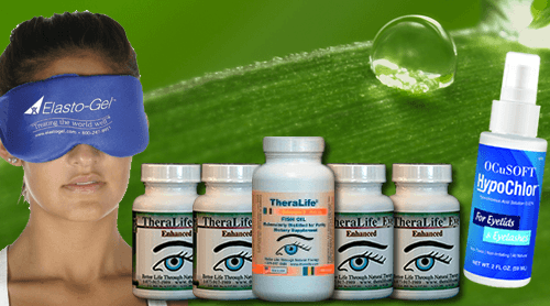
Why Theralife Works
- TheraLife Eye capsules – treat dry eyes from inside out, No more drops.
- Omega 3 fish oil – anti-inflammatory and provide lubrication for eye comfort
- Warm Compress – to unclog oil glands that cause chalazion, gently massage afterwards
- Eyelid Cleanser – to provide lid hygiene to prevent chalazion and blepharitis
Customer Story
No more recurring Chalazion
I was having painful chalazion just about every month. My eye doctor recommended cleaning my eye lids with baby shampoo, my lids are red and swollen, my vision became blurry, light sensitive. I found TheraLIfe on the internet and ordered the Chalazion Starter Kit. Withjn one week, my eye lids are no longer red and swollen, and I have not had any chalazion for the last 3 months. Dr. Yang works with me to make sure I am getting results. So happy to have found TheraLife.
D.Binder – Canada
Causes and treatments for crusty eyelashes
There are many possible causes for eyelashes that become crusty. Blepharitis, which is an inflammation of the eyelids, is one of the causes. It’s more common and can cause eyelashes to become dry and crusty. According to the American Academy of Ophthalmology (37% of patients have blepharitis at one time or another in their lives), To get rid of this problem, it is important to visit an eye care professional to have your eyes examined.
Blepharitis isn’t serious but it can be difficult to treat and recur. The infection is caused by a buildup of bacteria on the eyelid’s surface. In severe cases, blepharitis can cause pink eye or even rosacea. It’s important that you treat it as soon possible. If you experience any symptoms of this condition, it is important to consult your eye care provider.
When to consult a doctor about crusty eyelashes
Consult your doctor if your eyelashes are becoming dry and crusty. Allergies may trigger eczema. Your symptoms may be caused by a genetic or environmental factor. Your doctor may also be able to check your eye tear levels. You could also have seborrheic skin disease, which can cause flaky eyelids. This is caused due to a malfunctioning immune. The bacteria that live in the oil secretions on the eyelids are called malassezia.
How to treat crusty eyelashes
There are many ways to treat crusty eyelashes. The following are some of the recommended products by dermatologists for crusty eyelashes: Puriya Cream. You can keep your eyes clean and healthy by using a high quality cleanser or moisturizer. This is the best way to get rid of it.
If you have an infection that affects the eyelid, your doctor will want to investigate it to see if it’s a more serious condition. Blepharitis can be described as a condition that causes crusty eyes, dry eyes, and frequent blinking. In some cases, blepharitis could be the cause of your crusty, red eyelashes. Your doctor should be consulted to determine the cause of your crusty eyelashes.
Blepharitis, a more serious cause of crusty eyelashes and dilated eyes, is also possible. It is an infection affecting the conjunctiva, the thin tissue that covers the eye. It can affect either one or both eyes and is difficult to treat. Eyelid inflammation and redness can lead to eyelid tearing and infection. It is crucial to get treatment for Blepharitis as soon and as quickly as possible.
You can use a warm compress to alleviate chronic blepharitis. You should also avoid touching the eyelid with a cold compress. This will cause the crusts to swell and infected. To prevent bacteria spreading, you should avoid rubbing your eyes or using contact lenses with crusty eyelashes. A black teabag can be used as a warm compress. If you’re allergic to tea, steep it in boiling water and place it on your closed eyelid.
You can switch to natural cosmetics, if you are allergic or sensitive to certain ingredients. Natural products will not irritate eyes. Try to avoid makeup with perfumes. This can increase the chance of allergic reactions. It is also important that you clean your eyelids on a regular basis. You should wash your eyes with soap once a day or every other day. This will prevent you from developing these symptoms. You should consult a doctor if you have severe symptoms.
The most common cause of this condition is bacteria at or near the base. This condition causes red, crusty eyes. Some people also experience burning and itchy eyes. Those with blepharitis are more likely to suffer from other eye conditions. Before you attempt any cosmetic procedure, it is important to consult a doctor if you have blepharitis. If they do not have any other symptoms, they might be able to use topical creams or artificial tears.
Frequently Asked Questions
What happens if a chalazion is left untreated?
Chalazion left untreated can become a stye, which is infectious. It can also harden then you need surgery for removal. Get well now with TheraLife. Don’t delay
What triggers chalazion?
A clogged meibomian oil gland triggers the formation of a chalazion. This clogging is common amongst people who has chronic dry eyes.
How serious is a chalazion?
Conclusion
If your eyelashes seem dry and crusty it could be blepharitis. Flaky, itchy and other symptoms are caused by inflammation of your eyelids. Even if you don’t have blepharitis, it is worth seeking medical attention. It is not contagious, so don’t be alarmed if you’re experiencing this condition. You should consider an over-the-counter treatment if you have an eyelid stye.
References
2. Rodriguez-Garcia A, Loya-Garcia D, Hernandez-Quintela E, Navas A. Risk factors for ocular surface damage in Mexican patients with dry eye disease: a population-based study. Clin Ophthalmol. 2019;13:53-62. [PMC free article] [PubMed]
3. Choi FD, Juhasz MLW, Atanaskova Mesinkovska N. Topical ketoconazole: a systematic review of current dermatological applications and future developments. J Dermatolog Treat. 2019 Dec;30(8):760-771. [PubMed]
4. Ozkan J, Willcox MD. The Ocular Microbiome: Molecular Characterisation of a Unique and Low Microbial Environment. Curr Eye Res. 2019 Jul;44(7):685-694. [PubMed]
5. Khoo P, Ooi KG, Watson S. Effectiveness of pharmaceutical interventions for meibomian gland dysfunction: An evidence-based review of clinical trials. Clin Exp Ophthalmol. 2019 Jul;47(5):658-668. [PubMed]
6. Soh Qin R, Tong Hak Tien L. Healthcare delivery in meibomian gland dysfunction and blepharitis. Ocul Surf. 2019 Apr;17(2):176-178. [PubMed]
7. Fromstein SR, Harthan JS, Patel J, Opitz DL. Demodex blepharitis: clinical perspectives. Clin Optom (Auckl). 2018;10:57-63. [PMC free article] [PubMed]
8. Pflugfelder SC, Karpecki PM, Perez VL. Treatment of blepharitis: recent clinical trials. Ocul Surf. 2014 Oct;12(4):273-84. [PubMed]
9. Kanda Y, Kayama T, Okamoto S, Hashimoto M, Ishida C, Yanai T, Fukumoto M, Kunihiro E. Post-marketing surveillance of levofloxacin 0.5% ophthalmic solution for external ocular infections. Drugs R D. 2012 Dec 01;12(4):177-85. [PMC free article] [PubMed]
10. Veldman P, Colby K. Current evidence for topical azithromycin 1% ophthalmic solution in the treatment of blepharitis and blepharitis-associated ocular dryness. Int Ophthalmol Clin. 2011 Fall;51(4):43-52. [PubMed]
11. Hosseini K, Bourque LB, Hays RD. Development and evaluation of a measure of patient-reported symptoms of Blepharitis. Health Qual Life Outcomes. 2018 Jan 11;16(1):11. [PMC free article] [PubMed]


