Treat Your MGD with TheraLife
Root cause of MGD is chronic dry eyes. TheraLife has a comprehensive protocol that treats dry eyes, MGD, eliminates blepharitis and prevent watery eyes. Get help today
Call and talk to a doctor toll free 1-877-917-1989 US/Canada
Do you have MGD?
Meibomian Oil Glands (MG) are the glands located in the eyelids. because these glands provide protective oils, mucus, and proteins that keep the eyes moist and comfortable. However women, children, men across the nation find that they suffer from dry eye syndrome. This can be caused by blocked Meibomian glands ( Meibomian Gland Dysfunction – MGD).
Many patients undergo thermal pulsation or intense pulse heat treatments to unblock these glands,. However, there are better, less expensive solutions.
Younger Patients and Testing for MGD
Many people believe that dry eye syndrome is exclusively a problem for older patients. However, pilot studies and clinical evidence have shown that younger patients are vulnerable to dry eye disease and meibomian gland dysfunction (MGD). If this problem is not corrected, in time, this dysfunction can lead to glands dying.
Testing for MGD
Testing for dry eye disease was not readily available in the past. However, diagnostic testing for this disease is becoming more accessible to eye doctors. In addition, there are many diverse options available that allow for a wider range of measurements. The problem lies in the utilization in the pediatric patient population.
Children may not do well with invasive testing. Therefore, identifying any potential diseases in the eye becomes a frustrating process for patients and doctors alike. In order to assess the pediatric patient, specific dry eye diagnostic tests must be non-invasive and quick.
There are several options available: Meibography, Phenol red thread, and non-invasive Keratograph break-up time.
If a child is suspected of having dry eye disease, visualization of the meibomian glands is essential. Therefore, creating an image of the glands allows for current diagnosis. Meibography also provides a base for long-term observation of the glands in the future.
Imaging of pediatric glands, and adults who suffer critical to moderate advanced dry eye disease, is vital for diagnosis and treatment. The images obtained from Meibography are taken quickly and virtually painless for patients of all ages. One such imaging device is called “LipiView”
Comparable to traditional Schirmer’s testing to measure tear volume, PRT uses phenol red thread to obtain a measure of aqueous volume. The thread is placed at the lateral canthus (the outer edge of the eye) for 15 seconds.
During this time, the amount of fluid is measured in millimeters. The red thread changes color to yellow to show saturation. A normal result is 20 millimeters or greater. This is a preferred test because patients find little discomfort.
Non-invasite Keratograph break-up time (NIKBUT) for MGD Diagnosis
Many children are reluctant to having drops or dyes placed in their eyes. Unlike drops, dyes require additional patient cooperation once place on the eye surface. In order to accurately obtain results, the timing between dye placement and doctor observation must be precise. Equipment such as the Oculus Keratograph 5M requires patients to sit still for 15-20 seconds.
In younger patients, this may be difficult. The NIKBUT eliminates the use of dyes that many younger patients may find frightening or irritating. A Placido-disc topographer can also assist with the NIKBUT measurement by measuring the time between the eye-opening and the break-down of corneal fluids.
The next frontier is diagnostic testing is quickly becoming the pediatric dry eye assessment. Keenly in tune with making even more dry eye diagnoses in the adult population, clinicians are beginning to understand that diagnosing younger patients has many benefits.
With early detection of dry eye syndrome (DES), many of the problems that arise over time can be prevented, such as obstruction of the Meibomian glands and inflammation.
Finally, tests that once were considered invasive and almost impossible to perform on children are being redesigned with these younger patients in mind. As the old adage says, “An ounce of prevention is worth a pound of cure.” This is especially true in detecting dry eye disease in younger patients.
What to ask your eye doctor before signing up for heat treatments?
Before you spend thousands of dollars getting the heat treatment, ask your eye doctor to squeeze your Meibomian oil glands and tell you what comes out? Descriptions should be clear, cloudy, toothpaste or nothing comes out. If the secretions are thick, toothpaste like or nothing comes out. Heat treatments for MGD may not work.
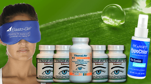
Get help from TheraLIfe
In order to keep the meibomian oil glands open, heat treatment is not enough. Get comprehensive relief for dry eyes, blepharitis, MGD at the same time plus heat treatment. Proven clinical success.

Stop MGD pain with TheraLIfe.
Frequently Asked Questions
How to unblock eye oil glands at home
Most popular method is to use warm compresses. However, if you have dry eyes, warm compress alone is not enough. Treat your dry eyes to control your MGD now.
How long does meibomian gland dysfunction last
MGD is a chronic condition that lasts a long time. As long as you have dry eyes, MGD will persist. If you seek heat treatments such as LipiFlow for IPL, the MGD will last from 6-9 months. Recommendation is to repeat treatment every 9 months.
Best eye drops for meibomian gland dysfunction
The eye drops to treat MGD are prescription drops for dry eyes. Examples are Restasis, Xiidra, and others. The gold standard to treat MGD is heat treatment.
How to unclog meibomian glands
Warm compress is the most popular method to unclog meibomian glands at home. However, you can go to the eye doctor’s office and express your meibomian glands to get the clogging out. For more severe cases, there is also intraductal meobomian probing – a surgical procedure.
Conclusion
MGD treatment algorithm in which treatment is added depending on the severity of MGD. The sequence of treatment addition is eyelid hygiene, eyelid warming and massage, artificial lubricants, topical azithromycin, topical emollient lubricant, oral tetracycline derivatives, lubricant ointment, and anti-inflammatory therapy. However, the efficacy of this treatment algorithm is yet to be evaluated. In addition to these treatments, new therapeutic modalities such as LipiView and IPL have emerged.
References
1. Bron AJ, Benjamin L, Snibson GR. Meibomian gland disease. Classification and grading of lid changes. Eye (Lond) 1991;5:395–411. doi: 10.1038/eye.1991.65.
2. Lekhanont K, Rojanaporn D, Chuck RS, Vongthongsri A. Prevalence of dry eye in Bangkok, Thailand. Cornea. 2006;25:1162–1167. doi: 10.1097/01.ico.0000244875.92879.1a.
3. Lin PY, Tsai SY, Cheng CY, Liu JH, Chou P, Hsu WM. Prevalence of dry eye among an elderly Chinese population in Taiwan: The Shihpai Eye Study. Ophthalmology. 2003;110:1096–1101. doi: 10.1016/S0161-6420(03)00262-8.
4. Uchino M, Dogru M, Yagi Y, Goto E, Tomita M, Kon T, Saiki M, Matsumoto Y, Uchino Y, Yokoi N, et al. The features of dry eye disease in a Japanese elderly population. Optom Vis Sci. 2006;83:797–802. doi: 10.1097/01.opx.0000232814.39651.fa.
5. Jie Y, Xu L, Wu YY, Jonas JB. Prevalence of dry eye among adult Chinese in the Beijing Eye Study. Eye (Lond) 2009;23:688–693. doi: 10.1038/sj.eye.6703101.
6. Tomlinson A, Bron AJ, Korb DR, Amano S, Paugh JR, Pearce EI, Yee R, Yokoi N, Arita R, Dogru M. The international workshop on meibomian gland dysfunction: Report of the diagnosis subcommittee. Invest Ophthalmol Vis Sci. 2011;52:2006–2049. doi: 10.1167/iovs.10-6997f.
7.
Dougherty JM, McCulley JP, Silvany RE, Meyer DR. The role of tetracycline in chronic blepharitis. Inhibition of lipase production in staphylococci. Invest Ophthalmol Vis Sci. 1991;32:2970–2975. [8. Tabbara KF, al-Kharashi SA, al-Mansouri SM, al-Omar OM, Cooper H, el-Asrar AM, Foulds G. Ocular levels of azithromycin. Arch Ophthalmol. 1998;116:1625–1628. doi: 10.1001/archopht.116.12.1625.
9. Schultz C. Safety and efficacy of cyclosporine in the treatment of chronic dry eye. Ophthalmol Eye Dis. 2014;6:37–42. doi: 10.4137/OED.S16067.
10. Ma X, Lu Y. Efficacy of intraductal meibomian gland probing on tear function in patients with obstructive meibomian gland dysfunction. Cornea. 2016;35:725–730. doi: 10.1097/ICO.0000000000000777.
11. Sik Sarman Z, Cucen B, Yuksel N, Cengiz A, Caglar Y. Effectiveness of intraductal meibomian gland probing for obstructive meibomian gland dysfunction. Cornea. 2016;35:721–724. doi: 10.1097/ICO.0000000000000820.
12.
Gumus K, Schuetzle KL, Pflugfelder SC. Randomized controlled crossover trial comparing the impact of sham or intranasal tear neurostimulation on conjunctival goblet cell degranulation. Am J Ophthalmol. 2017;177:159–168. doi: 10.1016/j.ajo.2017.03.002. [PMC free article] [PubMed] [CrossRef] [Google Scholar]13. Raulin C, Greve B, Grema H. IPL technology: A review. Lasers Surg Med. 2003;32:78–87. doi: 10.1002/lsm.10145.
14. Babilas P, Schreml S, Szeimies R-M, Landthaler M. Intense pulsed light (IPL): A review. Lasers Surg Med. 2010;42:93–104. doi: 10.1002/lsm.20877. [
15. Schuh A, Priglinger S, Messmer EM. Intense pulsed light (IPL) as a therapeutic option for Meibomian gland dysfunction. Ophthalmologe. 2019;116:982–988. doi: 10.1007/s00347-019-00955-z. (In German)
16. Brinton M, Kossler AL, Patel ZM, Loudin J, Franke M, Ta CN, Palanker D. Enhanced tearing by electrical stimulation of the anterior ethmoid nerve. Invest Ophthalmol Vis Sci. 2017;58:2341–2348. doi: 10.1167/iovs.16-21362.
17. Toyos R, McGill W, Briscoe D. Intense pulsed light treatment for dry eye disease due to meibomian gland dysfunction; a 3-year retrospective study. Photomed Laser Surg. 2015;33:41–46. doi: 10.1089/pho.2014.3819.
18. Dell SJ, Gaster RN, Barbarino SC, Cunningham DN. Prospective evaluation of intense pulsed light and meibomian gland expression efficacy on relieving signs and symptoms of dry eye disease due to meibomian gland dysfunction. Clin Ophthalmol. 2017;11:817–827. doi: 10.2147/OPTH.S130706.
19. Goldberg DJ. Current trends in intense pulsed light. J Clin Aesthet Dermatol. 2012;5:45–53.
20. Craig JP, Chen YH, Turnbull PR. Prospective trial of intense pulsed light for the treatment of meibomian gland dysfunction. Invest Ophthalmol Vis Sci. 2015;56:1965–1970. doi: 10.1167/iovs.14-15764. [
21. Arita R, Mizoguchi T, Fukuoka S, Morishige N. Multicenter study of intense pulsed light therapy for patients with refractory meibomian gland dysfunction. Cornea. 2018;37:1566–1571. doi: 10.1097/ICO.0000000000001687.
22. Arita R, Mizoguchi T, Fukuoka S, Morishige N. Therapeutic efficacy of intense pulsed light in patients with refractory meibomian gland dysfunction. Ocul Surf. 2019;17:104–110. doi: 10.1016/j.jtos.2018.11.004.
23. Yin Y, Liu N, Gong L, Song N. Changes in the meibomian gland after exposure to intense pulsed light in meibomian gland dysfunction (MGD) patients. Curr Eye Res. 2018;43:308–313. doi: 10.1080/02713683.2017.1406525.
24. Rong B, Tang Y, Tu P, Liu R, Qiao J, Song W, Toyos R, Yan X. Intense pulsed light applied directly on eyelids combined with meibomian gland expression to treat meibomian gland dysfunction. Photomed Laser Surg. 2018;36:326–332. doi: 10.1089/pho.2017.4402. [
25. Rong B, Tang Y, Liu R, Tu P, Qiao J, Song W, Yan X. Long-term effects of intense pulsed light combined with meibomian gland expression in the treatment of meibomian gland dysfunction. Photomed Laser Surg. 2018;36:562–567. doi: 10.1089/pho.2018.4499.
26. Jiang X, Lv H, Song H, Zhang M, Liu Y, Hu X, Li X, Wang W. Evaluation of the safety and effectiveness of intense pulsed light in the treatment of meibomian gland dysfunction. J Ophthalmol. 2016;2016(1910694) doi: 10.1155/2016/1910694.
27. Gupta PK, Vora GK, Matossian C, Kim M, Stinnett S. Outcomes of intense pulsed light therapy for treatment of evaporative dry eye disease. Can J Ophthalmol. 2016;51:249–253. doi: 10.1016/j.jcjo.2016.01.005.
28. Vegunta S, Patel D, Shen JF. Combination therapy of intense pulsed light therapy and meibomian gland expression (IPL/MGX) can improve dry eye symptoms and meibomian gland function in patients with refractory dry eye: A retrospective analysis. Cornea. 2016;35:318–322. doi: 10.1097/ICO.0000000000000735.
29. Karaca EE, Evren Kemer Ö, Özek D. Intense regulated pulse light for the meibomian gland dysfunction. Eur J Ophthalmol. 2020;30:289–292. doi: 10.1177/1120672118817687.
30. Seo KY, Kang SM, Ha DY, Chin HS, Jung JW. Long-term effects of intense pulsed light treatment on the ocular surface in patients with rosacea-associated meibomian gland dysfunction. Cont Lens Anterior Eye. 2018;41:430–435. doi: 10.1016/j.clae.2018.06.002.
31. Toyos R, Toyos M, Willcox J, Mulliniks H, Hoover J. Evaluation of the safety and efficacy of intense pulsed light treatment with meibomian gland expression of the upper eyelids for dry eye disease. Photobiomodul Photomed Laser Surg. 2019;37:527–531. doi: 10.1089/photob.2018.4599.
32. Moher D, Shamseer L, Clarke M, Ghersi D, Liberati A, Petticrew M, Shekelle P, Stewart LA. Preferred reporting items for systematic review and meta-analysis protocols (PRISMA-P) 2015 statement. Syst Rev. 2015;4(1) doi: 10.1186/2046-4053-4-1. PRISMA-P Group.
33.
Higgins J, Green S. Cochrane Handbook for systemic reviews of interventions. Version 5.1: The Cochrane Collaboration, 2011. https://handbook-5-1.cochrane.org. Accessed July 1, 2019.34. Vora GK, Gupta PK. Intense pulsed light therapy for the treatment of evaporative dry eye disease. Curr Opin Ophthalmol. 2015;26:314–318. doi: 10.1097/ICU.0000000000000166.
35. Albietz JM, Schmid KL. Intense pulsed light treatment and meibomian gland expression for moderate to advanced meibomian gland dysfunction. Clin Exp Optom. 2018;101:23–33. doi: 10.1111/cxo.12541.
36. Ngo W, SItu P, Keir N, Korb D, Blackie C, Simpson T. Psychometric properties and validation of the standard patient evaluation of eye dryness questionnaire. Cornea. 2013;32:1204–1210. doi: 10.1097/ICO.0b013e318294b0c0.
37. Vidas Pauk S, Petriček I, Jukić T, Popović-Suić S, Tomić M, Kalauz M, Jandroković S, Masnec S. Noninvasive tear film break-up time assessment using handheld lipid layer examination instrument. Acta Clin Croat. 2019;58:63–71. doi: 10.20471/acc.2019.58.01.09.


MGD is a common problem today and It can happen for a number of reasons. These are very important things to keep in mind for treating MGD. Thanks for sharing the informative article.
Harvey
Thank you. Share with your friends.