What happens during the pandemic
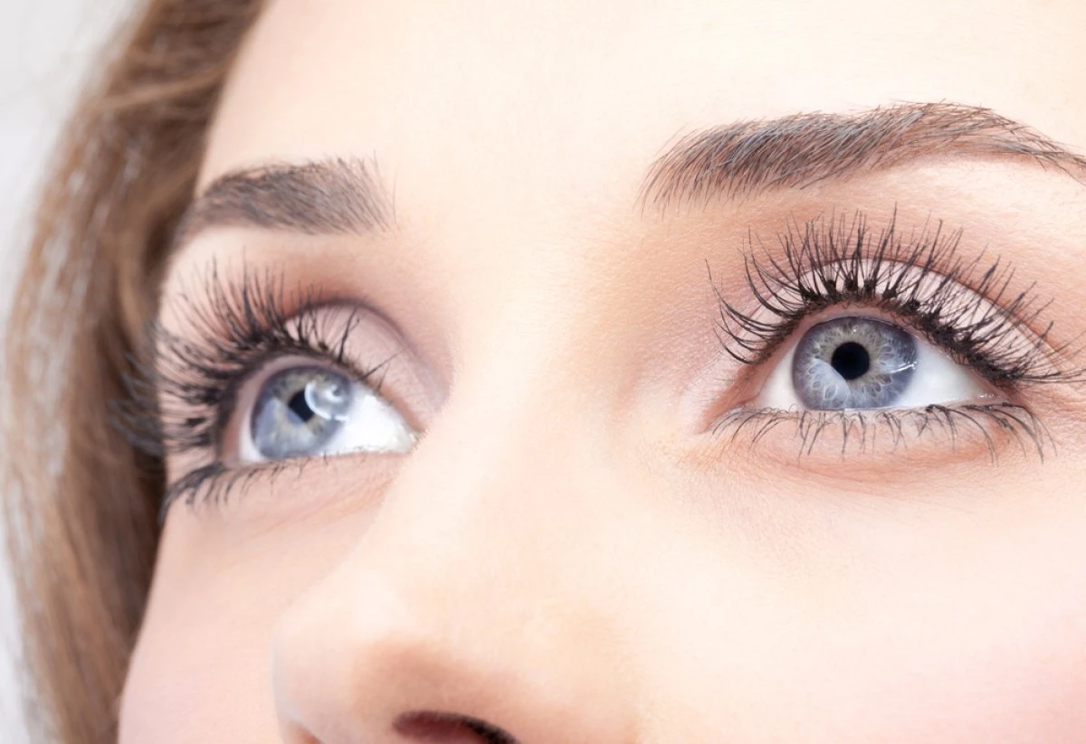
Dry eye gets worse during pandemic.
Everything about, well, everything in this pandemic is about “more.” More stress, more time, more visits to eye doctor’s offices but unable due to COVID-19. More problems to discuss and solve, perhaps online. Each problem that presents itself is more complex than it would have been in the new Before Covid.
And nothing has had more of “more” than Dry Eye Disease. More people are going to the eye doctor’s offices and being diagnosed with the disease for the first time.
Established Dry Eye Disease people arrive with more advanced diseases and more severe symptoms than they had before the pandemic. And it’s not just more surgical people presenting with Dry Eye Disease that need pre-op treatment. It’s EVERY surgical patient across the board needing to have treatment.
Why the dry eye increase?
In part, this is likely due to the multiplier effect on some of the well-understood causes. For example, increased screen time is a risk factor for developing and exacerbating the disease.
Whether they are working from home or simply filling the hours of a “shelter-at-home” day, people engage their screens at a rate 50-100% greater than pre-pandemic. All near tasks performed for more than a few minutes will reduce blink frequency. Screen use of any kind—desktop, laptop, tablet, or smartphone—will reduce blink amplitude.
Blink rate can be readily confirmed at the slit lamp and documented with LipiView, among other devices.
What happens to the Meibomian Oil Glands?
Without the “squeegee” effect of a complete blink, the meibomian glands cannot adequately evacuate meibum. The result is obstruction and, eventually, gland destruction. Increased evaporation of the tear film brought about by increased exposure time and reduced oil secretion leads to inflammation on the ocular surface. Inflammation reduces tear production even in the absence of systemic disease.
Medications can also cause dryness. Now we have the classic inflammation-driven vicious cycle of the disease in full bloom.
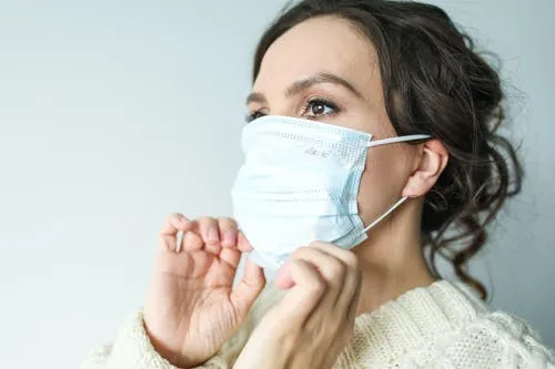
Mask Associated dry eye
Increased screen time alone seems insufficient to explain the dramatic increase seen in doctor’s offices.
In June 2020, there is a new entity that is called Mask-Associated Dry Eye or MADE.
Prolonged use of a mask is associated with more severe. At least a portion of the dryness generated by mask wear comes from the upward deflection of exhaled breath across the ocular surface. Air moving across a moist surface accelerates evaporation. In and of itself, this is sufficient to cause surface dryness and, of course, inflammation.
MADE may be more symptomatic than other diseases because evaporation causes cooling—temperature-sensitive nerves in the cornea cause pain.
Increase in Chalazion
We also see more meibomian gland disease and more chalazia than is typical.
Several hypotheses claim to explain this. Warm, moist air across the lower lid may optimize growing conditions for staph bacteria that are pro-inflammatory.
Experienced dry eye disease experts wonder if there has been an associated change in the local biome, both on the lids and the eye, resulting in more inflammation.
We see an increase in both the incidence and severity of Demodex infestation due to the micro-environmental changes caused by prolonged mask wear. Careful examination of eyelashes in people experiencing a significant increase in meibomian gland dysfunction (MGD) shows cylindrical dandruff, a finding typical for Demodex.
Let’s call it Mask-Associated Inflammatory Meibomitis from Demodex: MAIMeD.
Management of dry eye
With more severe disease and more symptomatic, we now face the challenge of treating it. Previously this was a challenge mostly met by specialists and those doctors who do cataract and refractive surgery.
Retinal and ophthalmic plastics eye doctors are also struggling. For example, reports indicate an increase in endophthalmitis cases post anti-VEGF injections – supposedly due to people wearing their masks during the injection.
How should we approach this phenomenon of “more”?
One approach is to be aggressive. Now is not the time to shy away from aggressive treatment of people with more aggressive forms of the disease, especially in the surgical setting. Previously we were able to control the disease by accurately gather pre-op data without making adjustments in standard scheduling formats. Now they may need to postpone surgery. Steroids are used aggressively in the pre-op setting. Higher potency steroids are used earlier and leaving on them longer.
No one foresees that people will stop wearing masks any time soon; now, eye doctors prescribe an immunomodulator (Cequa; Restasis; Xiidra). Once inflammation is present long-term anti-inflammatory therapy necessary.
Similarly, we have found that MGD previously managed with a mono-therapy approach now requires two or more concurrent interventions. Those offices with access to Intense Pulsed Light (IPL) have their units running all day, every day. Oral doxycycline, topical azithromycin long-term treatments are now required.
In summary
In short, in these days of “more” during the pandemic, we need to be more aggressive in all ways when diagnosing and treating the disease. More severe symptoms are bringing more people to doctor’s offices.
Peri-operative surgeries are seeing more severe disease. Eye doctors are treating more aggressively by all ophthalmic surgeons, including a greater willingness to use topical steroids in addition to long-term treatment with immunomodulators.
With the arrival of specific treatment options for Demodex on the horizon, it will help identify patients with more severe MGD who also harbor this infestation.
How TheraLife can help
TheraLife’s Eye formula targets to reduce inflammation, revive and restore normal tear functions intracellularly for relief. It is a patented technology that utilizes the mitochondria’s activation inside the cell to activate cellular functions.
Call and talk to a doctor toll-free 1-877-917-1989 US/Canada; International 650-949-6080
References
- The definition and classification of dry eye disease: report of the Definition and Classification Subcommittee of the International Dry Eye WorkShop. Ocul Surf. 2007;5:75–92
- Stern ME, Schaumburg CS, Pflugfelder SC. Dry eye as a mucosal autoimmune disease. Int Rev Immunol. 2013;32:19–41.
- Stevenson W, Chauhan SK, Dana R. Dry eye disease: an immune-mediated ocular surface disorder. Arch Ophthalmol. 2012;130:90–100.]
- Lemp MA, Crews LA, Bron AJ, Foulks GN, Sullivan BD. Distribution of aqueous-deficient and evaporative dry eye in a clinic-based patient cohort: a retrospective study. Cornea. 2012;31:472–478.
- Chia EM, Mitchell P, Rochtchina E, Lee AJ, Maroun R, Wang JJ. Prevalence and associations of dry eye syndrome in an older population: the Blue Mountains Eye Study. Clin Exp Ophthalmol. 2003;31:229–232.
- Schaumberg DA, Sullivan DA, Buring JE, Dana MR. Prevalence of dry eye syndrome among US women. Am J Ophthalmol. 2003;136:318–326.
- Ruprecht KW, Giere W, Wulle KG. Statistical contribution on symptomatic dry eye. Ophthalmologica. 1977;174:65–74.
- Bron AJ, Tomlinson A, Foulks GN, et al. Rethinking dry eEye disease: A perspective on clinical implications. Ocul Surf. 2014;12:1–31.
- Ridder WH, Zhang Y, Huang JF. Evaluation of reading speed and contrast sensitivity in dry eye disease. Optom Vis Sci. (3rd) 2013;90:37–44.
- Deschamps N, Ricaud X, Rabut G, Labbe A, Baudouin C, Denoyer A. The impact of dry eye disease on visual performance while driving. Am J Ophthalmol. 2013;156:184–189.
- Li M, Gong L, Chapin WJ, Zhu M. Assessment of vision-related quality of life in dry eye patients. Invest Ophthalmol Vis Sci. 2012;53:5722–5727.
- Schiffman RM, Walt JG, Jacobsen G, Doyle JJ, Lebovics G, Sumner W. Utility assessment among patients with dry eye disease. Ophthalmology. 2003;110:1412–1419.
- Labbe A, Wang YX, Jie Y, Baudouin C, Jonas JB, Xu L. Dry eye disease, dry eye symptoms and depression: the Beijing Eye Study. Br J Ophthalmol. 2013;97:1399–1403.
- Yu J, Asche CV, Fairchild CJ. The economic burden of dry eye disease in the United States: a decision tree analysis. Cornea. 2011;30:379–387.
- Foulks GN, Bron AJ. Meibomian gland dysfunction: a clinical scheme for description, diagnosis, classification, and grading. Ocul Surf. 2003;1:107–126.
- Management and therapy of dry eye disease: report of the Management and Therapy Subcommittee of the International Dry Eye WorkShop (2007) Ocul Surf. 2007;5:163–178.
- Sullivan BD, Crews LA, Messmer EM, et al. Correlations between commonly used objective signs and symptoms for the diagnosis of dry eye disease: clinical implications. Acta Ophthalmol. 2014;92:161–166.
- Methodologies to diagnose and monitor dry eye disease: Report of the Diagnostic Methodology Subcommittee of the International Dry Eye WorkShop (2007) Ocul Surf. 2007;5:108–152.
- Johnston PR, Rodriguez J, Lane KJ, Ousler G, Abelson MB. The interblink interval in normal and dry eye subjects. Clin Ophthalmol. 2013;7:253–259
- Arita R, Itoh K, Inoue K, Amano S. Noncontact infrared meibography to document age-related changes of the meibomian glands in a normal population. Ophthalmology. 2008;115:911–915.
- Nemeth J, Fodor E, Lang Z, et al. Lid-parallel conjunctival folds (LIPCOF) and dry eye: a multicentre study. Br J Ophthalmol. 2012;96:1380–1385.
- Höh H, Schirra F, Kienecker C, Ruprecht KW. Lid-parallel conjunctival folds are a sure diagnostic sign of dry eye. Ophthalmologe. 1995;92:802–808.