
Rheumatoid Arthritis Eye
Rheumatoid arthritis (RA) is an autoimmune disorder that attacks tendons and bones of joints.
Inflammations are common in other parts of our body, including skin, blood vessels. Typically this causes painful joints and stiffness.
Rheumatoid arthritis affect the eyes and can be a severe disease.
How TheraLife Can Help Relief Rheumatoid Arthritis Dry Eyes
What is rheumatoid arthritis (RA)
Rheumatological arthritis is a chronic autoimmune disorder that affects around 1% of American adults. The pathophysiology of RA involves chronic inflammation that causes proliferation of the synovial ducts and then cartilage destruction and bone erosion. It is the extra-articular manifestation of RA and is often the first symptom in the first stages. Rheumatopathologists must know how to treat rheumatic dermatitis and dry eyes.
While pain, swelling, and stiffness in the joints are the primary symptoms of RA, the inflammatory response of the misfiring immune system can cause a variety of other symptoms.
See more below.
Overlooked ocular manifestations
Despite rheumatism, RA usually presents synovitis in the fingers and wrists.
Ocular inflammatory conditions are commonly overlooked and largely underdiagnosed.
Usually present during the onset of RA population and occasionally represents the first sign. The sign of RA often affects the anterior part of the eye, keratoconjunctivitis.
Eye symptoms in RA
Symptoms associated with RA include dry eyes syndrome and episcleritis. Amongst vision problems resulting from retinal perforations are cornea and sclerotia, specifically scleromalacia perforans.
Multidisciplinary management is critical in cases involving peripheral ulcers and peripheral ulcer keratitis, which increase mortality.
Pathophysiology of ocular manifestations of RA
Pathophysiology Like the joints, the sclera and cornea contain proteoglycans and collagen. This histologic similarity likely accounts for many of the ocular manifestations of RA. The visual conditions associated with RA have pathological features in common with vasculitis – including vascular occlusion, infiltration, and fibrinoid necrosis.
Processes at play include immune complex deposition, secretion of collagenases.

TheraLife’s all-natural formula for rheumatoid arthritis eye and other chronic dry eye conditions targets the issue from the inside out. Try our products today.
Ocular manifestations of rheumatoid arthritis.
Several forms of eye disease can occur in patients with RA, and the clinical course of the ocular disease may be quite variable.
Several forms of eye disease can occur in patients with RA, and the clinical course of the ocular disease may be quite variable.
Extra-articular manifestations are also observed, including ophthalmic involvement.
Statistics
Ocular manifestations occur in 25% of patients with RA. The ocular manifestations include dry eye, episcleritis, keratitis, and keratoconjunctivitis sicca.
Keratoconjunctivitis sicca (dry eye) is RA’s most common ocular manifestation; it occurs in 15-25% of patients. The ocular manifestations include dry eye, episcleritis, keratitis, and keratoconjunctivitis sicca.
Episcleritis and scleritis occur less often-0.17 a % incidence of episcleritis with 0.67% incidence of scleritis. Ocular involvement, severe dry eye in particular.
Who is affected
Those with RA, relapsing polychondritis, and granulomatosis with polyangiitis (formerly known as Wegener’s granulomatosis)
Extra-articular involvement of organs in RA is significant. Ocular manifestations of RA are keratoconjunctivitis sicca, episcleritis, scleritis corneal changes, and retinal vasculitis. They were present in 27,2% of patients.
Women were more affected—the most common manifestation of ocular involvement was keratoconjunctivitis sicca.
Symptoms
The inflammatory condition of Rheumatoid Arthritis cause related eye symptoms.
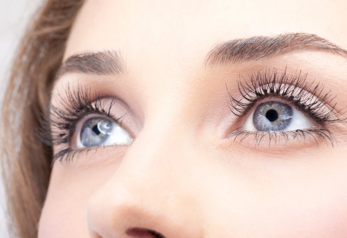
In addition to the common joint pain and inflammation experienced with rheumatoid arthritis, you may experience associated eye conditions, including eye redness, eye pain, eye dryness, blurred vision, Dry/gritty feelings, and sensitivity to light.
Redness that doesn’t go away with the use of over-the-counter eye drops (like Visine), severe pain (described as “deep, boring eye pain” by Dr. Lowder), light sensitivity, and perhaps reduced vision. RA can cause the eyewall (sclera), or the cornea, to become thin. Minor trauma could cause that part of the eyeball to split open.
Diagnosis
Diagnosis Patient’s complaints include red-eye and severe pain that awakens the patient from sleep. The eye pain can be described as a sensation of fullness to sharpness and stabbing in character.
Symptoms can include pain (sometimes severe), swelling, redness, blurred vision, tearing, and sensitivity to light. If you experience any of these issues, you should be evaluated by your ophthalmologist, And don’t delay seeking treatment. Time is of the essence to prevent eye damage and vision loss.
Diagnosis for dry eyes include tear break up time, Schirmer’s test, microscopic examinations of the eye using fluorescent dyes and more.
Treatments
If what’s going on with your eyes is more severe, your eye doctor may give you oral steroids and drugs that power down your immune system, like biologic medicines. You would get these if your eye problem comes from an overactive immune system, the same thing that causes RA.
Chronic topical or systemic steroids can also result in glaucoma.
In some cases, corticosteroid eye drops can control inflammation. Still, often the problem is too deep within the eye to be handled locally.
To learn more about Theralife’s treatment for dry eyes in RA.
Most common ocular manifestations
Dry eye syndrome
Dry eye syndrome, also known as keratoconjunctivitis sicca, is RA’s most common ocular manifestation. It can occur due to the meibomian gland, lacrimal gland, accessory lacrimal gland, or goblet cell dysfunction.
Vascular Scleritis
Rheumatoid arthritis (RA) is a systemic inflammatory disease associated with several extra-articular organ manifestations. Ocular manifestations involved with RA are keratoconjunctivitis sicca, episcleritis, scleritis, corneal changes, and retinal vasculitis.
The importance of the early diagnosis of ophthalmic disease in RA patients cannot be overemphasized since it permits the timely management of potentially sight-threatening severe complications. The presence of the ocular disease may also indicate ongoing membrane, bandage contact lenses, and tectonic, lamellar, or penetrating keratoplasty used to prevent and manage perforation.
Posterior scleritis
Posterior scleritis is characterized by the “T” sign on B-scan. This classic sign is produced by hyperreflectivity from the optic nerve and fluid under Tenon’s capsule.
Arthritis and your eyes. aao.org/eye-health/tips-prevention/arthritis-eyes-inflammation-steroids Ma W, et al. (2020). Study of factors influencing dry eye in rheumatoid
Orbital apex syndrome
Orbital apex syndrome (characterized by retro-orbital paralysis of extraocular muscles), impairment of the 1st division of the trigeminal nerve branches, and, frequently, extension to involve the optic nerve) resulting from orbital rheumatoid nodules has also been described.
Venous stasis retinopathy
As a rare complication, venous stasis retinopathy has been described in RA as part of a hyperviscosity syndrome secondary to polyclonal gammopathy.
Brown’s Syndrome
RA can cause Brown’s syndrome, restrictive strabismus due to scleritis without inflammation. This condition often presents without pain, despite its ability to lead to visual loss, astigmatism, and globe perforation.
A mild topical steroid 2 to 4 times per day can help treat the underlying inflammation often associated with dry eye. These are usually applied twice a day to treat the underlying inflammation.
Psoriatic Arthritis
In RA, ankylosing spondylitis and psoriatic arthritis increase the risk of eyeball inflammation. Taking oral or topical steroids also increases the risk. However, topical steroids can increase the risk of perforation and should be avoided.
Uveitis
RA inflames the uvea. That’s the layer of tissue between the back of your eye (the retina) and the sclera.
In addition to eye pain and light sensitivity, blurry vision is likely with uveitis.
Uveitis is inflammation of the uvea – the vascular layer of the eye, which is between the retina and the sclera.
Uveitis symptoms include pain, redness, blurred vision, and light sensitivity. If not controlled, uveitis can cause vision loss.
When does it become an emergency?
Urgent referral to an ophthalmologist with expertise in the management of ocular inflammation or cornea-external diseases of the eye. Symptoms such as a significant change in visual acuity for several days or weeks may represent severe corneal or scleral disease.
To read further –
Ocular Manifestations of Rheumatoid Arthritis: Implications of Recent Clinical Trials Manjeet S. Bhamra , 1 Irfan Gondal , 2 Abhimanyu Amarnani ,
Clinical management of ocular involvement in RA
Sicca- Dry Eye Disease
Keratoconjunctivitis, or Dry Eye Disease, is a common ophthalmic disorder primarily caused by reduced tear production and excessive liquid loss on the eye surface due to dry eyes. The evaporative dry eyes can coexist on a continuum in RA.
In severe RA, ocular involvement in the severe dry eye may exist independently from severe articular disease. Evaluate all patients with RA regardless of extra-ophthalmic manifestations.
A person with dry eyes might experience itching, a sand-like sensation in the eyes, light sensitivity, and redness. They may also notice a lack of moisture or tears in the eyes and blurred vision. Although many people turn to over-the-counter eye drops (artificial tears) for relief, the best treatment for dry eyes is prescription drops, which you can get from your ophthalmologist.
Learn more about TheraLife Dry Eye Treatment – all-natural and effective. No more eye drops.
RA is the most common systemic condition associated with scleritis. Ocular involvement, severe dry eye, in particular, may exist independently from powerful RA presentations. Regardless of whether ocular manifestations are present. Your doctor should evaluate the eyes of all patients with RA.
Chronic dry eye can cause corneal damage, leading to permanent vision loss. Your doctor will be able to provide the appropriate treatment for eye relief.
Dry eye and RA
Characteristics. Dry eye is the most common ophthalmic manifestation of RA.
Keratoconjunctivitis sicca (KCS) is RA’s most common ocular manifestation in 15-25% of patients.
Symptoms of dry eye syndrome include foreign body sensation, burning, decreased visual acuity, light sensitivity, and pruritus.
Treatment
You may get artificial tears, topical cyclosporine or ointment with steroids to ease inflammation. Punctal plugs into the tear duct to stop tear draining can help relieve dry eye pain.
Chronic dry eye can cause corneal damage, leading to permanent vision loss.
Learn more about RA dry eyes –
Ocular involvement in rheumatoid arthritis. aao.org/eyenet/article/ocular-involvement-in-rheumatoid-arthritis Tong L, et al. (2014)
Scleritis
RA is one of the main symptoms of scleritis characterized by vasodilation between superficial and deeper episcleral veins in both arteries.
Sclerosis is often a recurring condition when joint symptoms occur. It is sometimes caused by systemic vasculitis.
Scleritis occurs bilaterally, approximately 40% – 55% and can be classified as posterior or anterior depending on the relative position.
Scleric disorders.
Anterior sclerosis tends to become diffuse vascular sclerosis or nodule or necrotizing one.
Generally, it presents a severe ache amplified by eyes movement blurring, light sensitivity, and tears.
The presence of scleritis in patients with RA is associated with increased mortality. 1 Posterior scleritis can cause serous retinal detachment or chorioretinal folds. Other sequelae associated with scleritis include uveitis, cataracts, glaucoma, posterior segment damage, and peripheral ulcerative keratitis.
Treatment.
Topical medications are generally not effective in the treatment of scleritis. Therapy should manage the underlying RA with a multidisciplinary team approach involving the patient’s primary care physician and rheumatologist.
Oral NSAIDs are most effective in mild cases of scleritis, which usually affects multiple quadrants of the eye and presents with more severe symptoms such as discharge, photophobia, and decreased visual acuity.
Diagnosis
Typically, a diagnosis of episcleritis can be made clinically, and there are no published guidelines for establishing a diagnosis.
Peripheral ulcerative keratitis
RA patients often develop necrotizing scleritis or peripheral ulcerative keratitis.
Peripheral ulcerative keratitis (PUK) occurs in many systemic diseases, including RA. Signs of active PUK include guttering within the corneal limbus and corneal thinning. There is usually congestion and elevation of adjacent conjunctiva.
This occurs along with anterior scleritis but rarely with episcleritis.
Sclerosing keratitis occurs adjacent to the inflamed sclera and is characterized by corneal opacification and vascularization.
The scarring does not disappear with treatment and may progress with recurrences of the scleritis.
Anterior scleritis
Active scleritis involves inflammation of the deep episcleral layer associated with scleral edema. It tends to be bilateral in patients with RA. 14,15 Keratitis Corneal involvement often occurs with anterior scleritis but rarely with episcleritis.
Anterior scleritis can be nodular, diffuse, or necrotizing. It usually presents intense pain amplified by eye movements, blurry vision, photophobia, and tearing.
Posterior scleritis
Posterior scleritis or scleromalacia of cornea was not present in patients with the I or II stage of the disease. The new biotech therapies provide significant efficiency and safety. The evaluative disease activity requires intensive treatment with DMARDs, including biological treatment for rheumatoid arthritis and its ocular manifestations such as keratoconjunctivitis sicca and retinal vasculitis.
Treatment
For more severe diseases, the treatment of choice is oral corticosteroids (1 mg/kg/day of prednisone equivalents), with an optional bolus of methylprednisolone 1 g daily for three days. Depending on response and severity of scleritis at presentation, additional systemic medications include methotrexate, azathioprine.
Risks
The most vision-threatening sequelae include corneal perforation or ruptured globe from scleritis, particularly scleromalacia perforans. Multidisciplinary management is essential, especially in scleritis and peripheral ulcerative keratitis, associated with increased mortality in patients with RA.
It can present with pain, photophobia, tearing, and decreased visual acuity due to astigmatism or corneal opacity.
The differential diagnosis includes Mooren ulcer, Terrien marginal degeneration, herpetic keratitis, corneal ulcer secondary to dry eye, phlyctenular ulcers, and marginal keratitis.
Corneal perforation is a feared complication.
Episcleritis
Episcleritis is an inflammation of an episclera that lies beneath Tenon capsules. Symptoms presented with mild pain.
Scleritis RA is the most common cause of scleritis, which is an inflammation of the sclera characterized by vasodilation of the superficial and deep episcleral vessels.
Although the symptoms may be idiopathic, they are often linked to RA and some systemic diseases.
It occurs 40% bilaterally and appears with salmon-colored faces and light pain. Typically, they have sectoral distributions in diffuse patterns, diffuse patterns, or, less commonly, nodular forms. Contrary to scleritis, the eyes will disrupt with topical Phenylephrine.
Nodular episcleritis is confined to a limited portion of the episclera. In contrast, diffuse episcleritis involves a larger contiguous episcleral surface, affecting about 30% and 70% of the patients, respectively.
About 80% of all cases of episcleritis are limited to one eye, but in patients with systemic diseases such as RA, the incidence of bilateral eye involvement is higher. The overall incidence of episcleritis in the general population is less than 1/1000, with a female predominance [ 2 ]. The incidence of episcleritis in women between the ages of 45–64 was found to be 74 in 100,000, while the overall tendon sheath syndrome complicating rheumatoid arthritis.
Treatment.
Episcleritis diseases often become limited to themselves. Topical corticosteroids are also helpful in treating symptomatic inflammation.
Peripheral Ulcerative Keratitis (PUK)
PUK is an allergic condition characterized by the infiltration of immune complexes in vascular arcades of the cornea.
RA represents the most common disease, causing 34% of cases.
RA can also occur due to autoimmune infections or neurological conditions.
PUK usually comes with other ocular phases in severe RA and is secondary to scleritis, especially necrotizing scleritis.
PUK presents with stromal thinning and a secondary overlying epithelial defect and is characterized by neovascularization of the corneal periphery and crescent-shaped juxtalimbal ulcers.
Surgical considerations in RA Ocular Disease
Surgical trauma can trigger a macrophage and a neutrophil infiltration-causing progressively immunologic reaction and formation of immune complexes. These immune complexes can also crystals called cryo-precipitates.
The immuno-complementary proteins are deposited in the cornea and are responsible for the keratotic reaction. During the initial inflammation, fibroblasts produce complement components C1 and contribute to the activation of the classical complement cascade.
Surgery may interfere with the eye’s immune privilege or cause inflammation by the immune reaction through the cellular mimics.
Routine Eye Health Exam
People with RA should also visit an ophthalmologist if they experience eye symptoms, such as itching, foreign body sensation, redness, pain, or vision loss.
Finally, it’s important to note that other conditions like acne and rosacea can lead to eye problems.
See more about ocular rosacea.
It’s good for people with RA to be aware of this, although it’s still critical to see your doctor.
What is rheumatoid arthritis?
Rheumatoid arthritis causes inflammation and is a systemic disease affecting the entire body.
Rheumatoid Arthritis (RA) is a chronic systemic autoimmune disease that affects approximately 1% of the adult U.S. population. The pathophysiology of RA involves chronic inflammation leading to a proliferation of the synovium with subsequent cartilage damage and bony erosions.
Ocular inflammation is an extra-articular manifestation of RA.
RA is the most common form of autoimmune disease.
It affects more than 1.3 million Americans.
About 75% of RA patients are women. 1–3% of women may get rheumatoid arthritis in their lifetime. The disease most often begins between the ages of 30 and 50. However, RA can start at any age.
RA is a chronic disease that causes joint pain, stiffness, swelling, and decreased movement of the joints. Small joints in the hands and feet are most commonly affected.
Sometimes RA can affect other organs, such as the eyes, skin, or lungs.
In addition to causing joint problems, rheumatoid arthritis can often affect other body organs, such as the skin, eyes, lungs, and blood vessels.
The joint stiffness in active RA is often the worst in the morning. It may last one to two hours (or even the whole day). It generally improves with the movement of the joints. Stiffness for a long time in the morning is a clue that you may have RA, as this is not common in other conditions. For instance, osteoarthritis most often does not cause prolonged morning stiffness.
Other signs and symptoms that can occur in RA include:
- Loss of energy
- Low-grade fevers
- Loss of appetite
- Firm lumps called rheumatoid nodules grow beneath the skin in places such as the elbow and hands.
Causes
RA is an autoimmune disease. Your immune system is supposed to attack foreigners in your body, like bacteria and viruses, by creating inflammation.
The immune system mistakenly sends inflammation to your healthy tissue in an autoimmune disease.
The immune system creates a lot of inflammation sent to your joints, causing pain and swelling. If the inflammation remains present for an extended period, it can cause damage to the joint. This damage typically cannot be reversed once it occurs.
The exact cause of RA is not known. There is evidence that autoimmune conditions run in families. For instance, specific genes you are born with may make you more likely to get RA.
Smoking is also a known risk factor for causing RA.
Diagnosis
Diagnosis for rheumatoid arthritis can be determined from joint involvement, symptoms duration, and serum studies. Urine and liver, and blood test results may help identify other symptoms or guide the doses of medication. A joint MRI scan and total blood counts were also included in this study. Differential C-reactive protein levels and erythrocyte sedimentation rate were also measured.
RA is diagnosed by examining blood test results, examining the joints and organs, and reviewing x-ray or ultrasound images.
There is no single test to diagnose RA.
Blood tests are run to look for antibodies in the blood that can be seen in RA.
Antibodies are small proteins in the bloodstream that help fight against foreign substances called antigens. Sometimes these antibodies are found in people without RA.
Blood tests are also run to look for high levels of inflammation.
The symptoms of RA can be very mild, making the diagnosis more difficult.
Some viral infections can cause symptoms that can be mistaken for RA. A rheumatologist is a physician with the skill and knowledge to reach a correct diagnosis of RA and recommend a treatment plan.
Abnormal blood tests commonly seen in RA include:
- Anemia (a low red blood cell count)
- Rheumatoid factor (an antibody, or blood protein, found in about 80% of patients with RA in time but as few as 30% at the start of arthritis)
- Antibodies to cyclic citrullinated peptides (pieces of proteins), or anti-CCP for short (found in 60 – 70% of patients with RA)
- Elevated erythrocyte sedimentation rate (a blood test that, in most patients with RA, confirms the amount of inflammation in the joints)
X-rays can help in detecting RA but may be expected in early arthritis. Even if normal, initial X-rays may be helpful later to show if the disease is progressing. MRI and ultrasound scanning can help confirm or judge the severity of RA.
RA is chronic arthritis. Generally, the symptoms will need to be present for more than three months to consider this diagnosis. However, some patients are diagnosed sooner.
Treatment
Therapy for RA has significantly improved in the past 30 years.
Current treatments give most patients good or excellent relief of symptoms and let them keep functioning at, or near, normal levels.
Many patients can have no signs of active disease with the proper medications. When the symptoms are completely controlled, the condition is in “remission.”
There is no cure for RA.
The goal of treatment is to improve your joint pain and swelling and improve your ability to perform day-to-day activities.
Starting medication as soon as possible helps prevent your joints from having lasting or possibly permanent damage.
No single treatment works for all patients. Many people with RA must change their treatment at least once.
RA patients should begin their treatment with disease-modifying antirheumatic drugs (DMARDs). These drugs relieve symptoms and slow the progression of joint damage. Doctors often prescribe DMARDs, nonsteroidal anti-inflammatory drugs, NSAIDs, and low-dose corticosteroids to decrease swelling and pain.
DMARDs have greatly improved the pain, swelling, and quality of life for nearly all patients with RA. Ask your rheumatologist about the need for DMARD therapy and the risks and benefits of these drugs.
Common DMARDs include methotrexate (Rheumatrex, Trexall, Otrexup, Rasuvo), leflunomide (Arava), hydroxychloroquine (Plaquenil) and sulfasalazine (Azulfidine). Azathioprine and Cyclosporine can occasionally be used but not as frequently as other drugs work more efficiently.
Patients with more severe diseases may need medications called biologic response modifiers or “biologic agents.” They can block immune system chemical signals that lead to inflammation and joint/tissue damage. FDA-approved drugs of this type include abatacept (Orencia), adalimumab (Humira), anakinra (Kineret), certolizumab (Cimzia), etanercept (Enbrel), golimumab (Simponi) infliximab (Remicade), rituximab (Rituxan, MabThera), sarilumab (Kevzara) and tocilizumab (Actemra). Patients often take these drugs with methotrexate, as the mix of medicines is more helpful.
Janus kinase (JAK) inhibitors are another type of DMARD. People who cannot be treated with methotrexate alone. Co-administer with a JAK inhibitor such as tofacitinib (Xeljanz) or baracitinib (Olumiant).
The best treatment of RA needs more than medicines alone.
Patient education, such as coping with RA, is also essential.
Proper care often requires a team of providers, including rheumatologists, primary care physicians, and physical and occupational therapists.
You will need frequent visits with your rheumatologist.
These checkups let your doctor track the course of your disease and check for any side effects of your medications.
Also, you likely will need to repeat blood tests and X-rays or ultrasounds from time to time.
Living with RA
It is essential to be physically active most of the time but to sometimes scale back activities when the disease flares.
Rest is helpful when a joint is inflamed or when you feel tired. At these times, do gentle range-of-motion exercises, such as stretching. This will keep the joint flexible.
RA patients are encouraged to do low-impact aerobic exercises, such as walking, to boost muscle strength when they feel better. This will improve your overall health and lower the pressure on your joints. A physical or occupational therapist can help you find which activities are best for you and what level or pace you should do them.
Finding that you have a chronic illness is a life-changing event. It can cause worry and sometimes feelings of isolation or depression. Thanks to greatly improved treatments, these feelings tend to decrease with time as energy improves and pain and stiffness decrease.
Discuss these normal feelings with your health care providers. They can provide helpful information and resources.
Frequently Asked Questions
What are the symptoms of arthritis in the eye?
Symptoms of arthritis in the eye include dryness, inflammation, eye pain, light sensitivity, blurry vision, watery eyes, and blepharitis, MGD. Consult your eye doctor for treatment. Avoid use of steroids which can result in glaucoma.
What eye condition is associated with arthritis?
What eye condition is associated with arthritis?
Conclusion
Rheumatoid arthritis can lead to serious eye problems that should not be ignored. In order to treat this condition, you need an eye specialist- called Opthalmology. Consult with TheraLIfe and get arthritis dry eye under control with an all natural remedy that works.
References
1. Felson DT, Anderson JJ, Boers M. American College of Rheumatology. Preliminary definition of improvement in rheumatoid arthritis. Arthritis Rheum. 1995;38:727–735.
2. Turesson C, O’Fallon WM, Crowson CS, Gabriel SE, Matteson EL. Extra-articular disease manifestations in rheumatoid arthritis: incidence trends and risk factors over 46 years. Ann. Rheum. Dis. 2003;62:722–727.
3. Sahatçiu-Meka V, Rexhepi S, Manxhuka-Kerliu S, Rexhepi M. Extraaricular manifestation of seronegative and seropositive reumathoid arthritis. Bosn. J Basic Med Sci. 2010;10(1):27–31.
4. Lilleby V, Gran JT. Systemic rheumatoid arthritis. Tidsskr Nor Laegeforen. 1997;117(29):4223–4225.
5. Goronzy JJ, Weyand CM. Developments in the scientific under-standing of rheumatoid arthritis. Arthritis Res Ther. 2009;11(5):249.
6. Cimmino MA, Salvarani C, Macchioni P, et al. Extra-articular manifestations in YZ Italian patients with rheumatoid arthritis. Rheumatol Int. 2000;19(6):213–217.
7. Kanski JJ. The eye in systemic disease. London: Butterworts; 1986.
8. Whitson WE, Krachmer JH. Adult rheumatoid arthritis. In: Gold DH, Weingeist TA, editors. The eye in systemic disease. Philadelphia: JB Lippincott; 1990. pp. 127–208.
9. Hakin KN, Ham J, Lightman SL. Use of orbital floor steroids in the management of patients with uniocular non-necrotizing scleritis. Br J Ophthalmol. 1991;75(6):337–339.
10. Galor A, Jabs DA, Leder HA, Kedhar SR, Dunn JP, Peters GB. Comparison of antimetabolite drugs as corticosteroid-sparing therapy for noninfectious ocular inflammation. Ophthalmol. 2008;115(10):1826–1832.
11. Arnett FC, Edworthy SM, Bloch DA. The American Rheumatism Association 1987 revised criteria for the classification of rheumatoid arthritis. Arthritis Rheum. 1988;31:315–324.
12. Goto E, Matsumoto Y, Kamoi M, Endo K, Ishida R, Dogru M. Tear evaporation rates in Sjögren syndrome and non-Sjögren dry eye patients. Am J Ophthalmol. 2007;144(1):81–85.
13. Barr CC, Davis H, Culberson WW. Rheumatoid scleritis. Ophthalmol. 1981;88:1269–1273.
14. Hori Y, Maeda N, Sakamoto M, Koh S, Inoue T, Tano Y. Bacteriologic profile of the conjunctiva in the patients with dry eye. Am J Ophthalmol. 2008;146(5):729–734.
15. Sobrin L, Kim EC, Christen W, Papadaki T, Letko E, Foster CS. Infliximab therapy for the treatment of refractory ocular infiammatory disease. Arch Ophthalmol. 2007;125(7):895–900.
16. Sainz de la Maza M, Foster CS, Jabbur NS. Scleritis associated with rheumatoid arthritis and with other systemic immune-mediated diseases. Ophthalmol. 1994;101(7):1281–1286.
17. Lin CP, Shih MH, Su CY. Scleritis. Surv Ophthalmol. 2006;51(3):288–289.
18. Fong LP, Sainz de la Maza M, Rice BA, Kupferman AE, Foster CS. Immunopathology of scleritis Ophthalmol . 1991;98(4):472–479.
19. Sainz de la Maza M, Jabbur NS, Foster CS. Severity of scleritis and episcleritis. Ophthalmol. 1994;101(2):389–396.
20. Wakefield D, McCluskey P. Cyclosporin therapy for severe scleritis. Br J Ophthalmol. 1989;73(9):743–746.
21. Cazabon S, Over K, Butcher J. The successful use of infliximab in resistant relapsing polychondritis and associated scleritis. Eye. 2005;19(2):222–224.
22. Murphy CC, Ayliffe WH, Booth A, Makanjuola D, Andrews PA, Jayne D. Tumor necrosis factor alpha blockade with infliximab for refractory uveitis and scleritis. Ophthalmol. 2004;111(2):352–356.
23. Sobrin L, Kim EC, Christen W, Papadaki T, Letko E, Foster CS. Infliximab therapy for the treatment of refractory ocular inflammatory disease. Arch Ophthalmol. 2007;125(7):895–900.
24. Ahmadi-Simab K, Lamprecht P, Nolle B, Ai M, Gross WL. Successful treatment of refractory anterior scleritis in primary Sjogren’s syndrome with rituximab. Ann Rheum Dis. 2005;64(7):1087–1088.
25. Lim L, Suhler EB, Smith JR. Biologic therapies for inflammatory eye disease. Clin Exp Ophthalmol. 2006;34(4):365–374.
26. Sobrin L, Christen W, Foster CS. Mycophenolate mofetil after methotrexate failure or intolerance in the treatment of scleritis and uveitis. Ophthalmol. 2008;115(8):1416–1421.
27. Papaliodis GN, Chu D, Foster CS. Treatment of ocular inflammatory disorders with daclizumab. Ophthalmol. 2003;110(4):786–789.
28. Sokolović Š, Kasumagić Š, MarkićĐurović M, AganovićMušinović I. The impact of Rituximab therapy on the chromosomes of patients with rheumatoid arthritis. Bosn J Basic Med Sci. 2010;10(2):121–124.