
This article deals with the perceptions of eyecare providers regarding the clinical management of dry eye.
The survey is taken from community optometrists, comprehensive ophthalmologists, and cornea specialists.
What are the most common dry eye symptoms at doctor’s offices?
The following are dry eye symptoms most often presented to an eye doctor.
Burning- 47%
Foreign body sensation – 30%
Tearing – 17%
What are the most common dry eye treatments?
Artificial tears, even though eye doctors noted high failure rates, even with Restasis (cyclosporine – A.
Co Morbilities for Dry Eye Disease
- Rheumatoid arthritis,
- Sjögren syndrome – dry eyes and dry mouth,
- anxiety and depression,
- history of photorefractive surgery – LASIK surgery
- smoking,
- thyroid disease -Graves Disease, Hypo, and hyperthyroidism.
Eye doctors relied more on patient history in guiding their clinical decisions than objective signs.
The survey underscores the inconsistency when comparing subjective symptoms with objective signs.
There is an urgent need for the development of reliable metrics to quantify better dry eye symptoms. We need a more sensitive and specific test that can be used as the gold standard to diagnose dry eye.
What is dry eye disease?
Dry Eye Disease, also known as Dry Eye Syndrome or Keratoconjunctivitis Sicca, is a clinical diagnosis determined by your eye doctor.
Since many possible factors or underlying causes, complete health history and thorough eye examination are necessary to diagnose and treat dry eye conditions properly.
Your eye doctor may have you complete a symptom questionnaire throughout Dry Eye Management to assess the effectiveness of the current treatment or adjust as needed.
What are the dry eye tests?
There are various tests your eye doctor may perform to measure the severity of dry eye you are experiencing. At Eyes For Life, your doctor may choose one or more of the following evaluations:
- LipiScan™
- Meibomian Gland Evaluator™
- SPEED™Questionnaire
- InflammaDry
- TearLab™ Osmolarity
- Tear Breakup Time
- Epithelial Staining
- The Schirmer Test
- The Phenol Red Thread Test
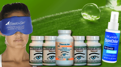
How can TheraLife help?
TheraLife Eye capsules target to normalize tear production glands from inside out. It is an oral dry eye treatment system, not eye drops.
References
1. Nichols KK, Nichols JJ, Mitchen GL. The lack of association between signs and symptoms in patients with dry eye disease. Cornea. 2004;23(8):762–770.
2. Schirmer O. Studien zur physiologie und pathologie der tranen-absonderung und tranenabfuhr. Graefes Arch Clin Exp Ophthalmol. 1903;56:197–291. German.
3. De Roetth A. Lacrimation in normal eyes. Arch Ophthalmol. 1953;49(2):185–189. ]
4. Lemp MA. Report of the National Eye Institute/Industry workshop on the Clinical Trials in Dry Eyes. CLAO J. 1995;21(4):221–232.
5. Dry Eye Workshop. The definition and classification of dry eye disease: report of the definition and classification subcommittee of the International Dry Eye Workshop. Ocul Surf. 2007;5(2):75–92. [No authors listed]
6. Hashemi H, Khabazkhoob M, Kheirkhah A, et al. Prevalence of dry eye syndrome in an adult population. Clin Experiment Ophthalmol. 2013 Aug 8; [Epub ahead of print]
7. Scuderi G, Contestabile MT, Gagliano C, Iacovello D, Scuderi L, Avitabile T. Effects of phytoestrogen in postmenopausal women with dry eye syndrome: a randomized clinical trial. Can J Ophthalmol. 2012;47(6):489–492.
8. Rocha EM, Mantelli F, Nominato, Bonini S. Hormones and dry eye syndrome: an update on what we do and don’t know. Curr Opin Ophthalmol. 2013;24(4):348–355. [
9. Wong J, Lan W, Ong LM, Tong L. Non-hormonal systemic medications and dry eye. Ocul Surf. 2011;9(4):212–226. [
10. Farris RL. The dry eye: its mechanism and therapy, with evidence that contact lens is a cause. CLAO J. 1986;12(4):234–246.
11. Grus FH, Sabuncuo P, Augustin A, Pfeiffer N. Effect of smoking on tear proteins. Graefes Arch Clin Exp Ophthalmol. 2002;240(11):889–892
12. Quinto GG, Camacho W, Behrens A. Postrefractive surgery dry eye. Curr Opin Ophthalmol. 2008;19(4):335–341.
13. Moschos MM, Chatziralli IP, Siasou G, Papazisis L. Visual problems in young adults due to computer use. Klin Monbl Augenheilkd. 2012;229(4):379–381.German.
14. Abusharha AA, Pearce EI. The effect of low humidity on the human tear film. Cornea. 2013;32(4):429–434.
15. Palmowski AM, Ruprecht KW. The cornea and systemic diseases. Curr Opin Ophthalmol. 1995;6(4):17–20.
16. Shapiro A, Merin S. Schirmer test and break-up time of tear film in normal subjects. Am J Ophthalmol. 1979;88(4):752–757.
17. Jordan A, Baum J. Basic tear flow. Does it exist? Ophthalmology. 1980;87:920–930.
18. Li N, Deng XG, He MF. Comparison of the Schirmer I test with and without topical anesthesia for diagnosing dry eye. Int J Ophthalmol. 2012;5(4):478–481
19. Serin D, Karsloglu S, Kyan A, Alagoz G. A simple approach to the repeatability of the Schirmer test without anesthesia: eyes open or closed. Cornea. 2007;26(8):903–906
20. Kashkouli MB, Pakdel F, Amani A, Asefi M, Aghai GH, Falavarjani KG. A modified Schirmer test in dry eye and normal subjects: open versus closed eye and 1-minute versus 5-minutes tests. Cornea. 2010;29(4):384–387
21. Nichols KK, Mitchell GL, Zadnik K. The repeatability of clinical measurements of dry eye. Cornea. 2004;23(3):272–285.
22. Bitton E, Wittich W. Influence of eye position on the Schirmer tear test. Cont Lens Anterior Eye. 2013 Dec 18; [Epub ahead of print]
23. Feldman F, Wood MM. Evaluation of the Schirmer tear test. Can J Ophthalmol. 1979;14(4):257–259.
24. Yoon KC, Im SK, Kim HG, You IC. Usefulness of double vital staining with 1% fluorescein and 1% lissamine green in patients with dry eye syndrome. Cornea. 2011;30(9):972–976.
25. Savini G, Barboni P, Zanini M. The incidence and risk factors for developing dry eye after myopic LASIK. Am J Ophthalmol. 2006;142(2):355–356
26. Feenstra RP, Tseng SC. What is actually stained by rose bengal? Arch Ophthalmol. 1992;110(7):984–993]
27. Schein OD, Tielsch JM, Munõz B, Bandeen-Roche K, West S. Relation between signs and symptoms of dry eye in the elderly: a population-based perspective. Ophthalmology. 1997;104(9):1395–1401
28. Khan-Lim D, Berry M. Still confused about rose bengal? Curr Eye Res. 2004;29(4–5):311–317
29. Kim J, Foulks GN. Evaluation of the effect of lissamine green and rose bengal on human corneal epitheliual cells. Cornea. 1999;18(3):328–332
30. Machado LM, Castro RS, Fontes BM. Staining patterns in dry eye syndrome: rose bengal versus lissamine green. Cornea. 2009;28(7):732–734.
31. Hamrah P, Alipour F, Jiang S, Sohn JH, Foulks GN. Optimizing evaluation of lissamine green parameters for ocular surface staining. Eye (Lond) 2011;25(11):1429–1434.
32. Berntsen DA, Mitchell GL, Nichols JJ. Reliability of grading lissamine green conjunctival staining. Cornea. 2006;25(6):695–700.
33. Bron AJ, Evans VE, Smith JA. Grading of corneal and conjunctival staining in the context of other dry eye tests. Cornea. 2003;22(7):640–650
34. Van Bijsterveld OP. Diagnostic tests in the sicca syndrome. Arch Ophthalmol. 1969;82:10–14
35. Savini G, Prabhawasat P, Kojima T, Grueterich M, Espana E, Goto E. The challenge of dry eye diagnosis. Clin Ophthalmol. 2008;2(1):31–55
36. Xu KP, Yagi Y, Toda I, Tsubota K. Tear function index: a new measure of dry eye. Arch Ophthalmol. 1995;113(1):84–88
37. Kaye SB, Sims G, Willoughby C, Field AE, Longman L, Brown MC. Modification of the tear function index and its use in the diagnosis of Sjogren’s syndrome. Br J Ophthalmol. 2001;85(2):193–199
38. Prabhasawat P, Tseng SC. Frequent association of delayed tear clearance in ocular irritation. Br J Ophthalmol. 1998;82(6):666–675
39. Afonso AA, Monroy D, Stern ME, Feuer WJ, Tseng SC, Pflugfelder SC. Correlation of tear fluorescein clearance and Schirmer test scores with ocular irritation symptoms. Ophthalmology. 1999;106(4):803–810.
40. Lemp MA. Breakup of the tear film. Int Ophthalmol Clin. 1973;13(1):97–102.
41. Lemp MA, Hamill JR. Factors affecting tear film breakup in normal eyes. Arch Ophthalmol. 1973;89(2):103–105.
42. Norn MS. Desiccation of the precorneal film. I. Cornea wetting-time. Acta Ophthalmol (Copenh) 1969;47(4):865–880.
43. Norn MS. Desiccation of the precorneal film. II. Permanent discontinuity and dellen. Acta Ophthalmol (Copenh) 1969;47(4):881–889.
44. Vanley GT, Leopold IH, Gregg TH. Interpretation of tear film breakup. Arch Ophthalmol. 1977;95(3):445–448.
45. Lin YY, Carrel H, Wang IJ, et al. Effect of tear film break-up on higher order aberrations of the anterior cornea in normal, dry, and post-LASIK eyes. J Refract Surg. 2005;21(5):S525–S529.
46. Tsubota K, Nakamori K. Dry eyes and video display terminals. N Engl J Med. 1993;328(8):584
47. Rieger G. The importance of the precorneal tear film for the quality of optical imaging. Br J Ophthalmol. 1992;76(3):157–158
48. Goto E, Yagi Y, Matsumoto Y, et al. Impaired functional visual acuity of dry eye patients. Am J Ophthalmol. 2002;133(2):181–186. [
49. Ishida R, Kojima T, Dogru R, et al. The application of a new continuous functional visual acuity measurement system in dry eye syndromes. Am J Ophthalmol. 2005;139(2):253–258.
50. Kaido M, Dogru M, Ishida R, Tsubota K. Concept of functional visual acuity and its application. Cornea. 2007;26(9 Suppl 1):S29–S35.
51. Mainstone JC, Bruce AS, Golding TR. Tear meniscus measurement in the diagnosis of dry eye. Curr Eye Res. 1996;15(6):653–661.
52. Guillon JP. Non-invasive Tearscope Plus routine for contact lens fitting. Cont Lens Anterior Eye. 1998;21(Suppl 1):S31–S40.
53. Fodor E, Hagyó K, Resch M, Somodi D, Németh J. Comparison of Tearscope Plus versus slit lamp measurements of the inferior tear meniscus height in normal individuals. Eur J Ophthalmol. 2010;20(5):819–824.
54. Lamberts DW, Foster CS, Perry HD. Schirmer test after topical anesthesia and the tear meniscus height in normal eyes. Arch Ophthalmol. 1979;97(6):1082–1085.
55. Miller WL, Doughty MJ, Narayanan S, et al. A comparison of tear volume (by tear meniscus height and phenol red thread test) and tear fluid osmolality measures in non-lens wearers and in contact lens wearers. Eye Contact Lens. 2004;30(3):132–137.
56. Savini G, Barboni P, Zanini M. Tear meniscus evaluation by optical coherence tomography. Ophthalmic Surg Lasers Imaging. 2006;37(2):112–118
57. Yokoi N, Bron A, Tiffany J, Brown N, Hsuan J, Fowler C. Reflective meniscometry: a non-invasive method to measure tear meniscus curvature. Br J Ophthalmol. 1999;83(1):92–97.
58. Bandlitz S, Purslow C, Murphy PJ, Pult H. Comparison of a new portable digital meniscometer and optical coherence tomography in tear meniscus radius measurement. Acta Ophthalmol. 2013 Oct 7; [Epub ahead of print]
59. Bandlitz S, Purslow C, Murphy PJ, Pult H, Bron AJ. A new portable digital meniscometer. Optom Vis Sci. 2014;91(1):e1–e8.
60. Chen F, Shen M, Chen W, et al. Tear meniscus volume in dry eye after punctal occlusion. Invest Ophthalmol Vis Sci. 2010;51(4):1965–1969
61. Nguyen P, Huang D, Li Y, et al. Correlation between optical coherence tomography-derived assessments of lower tear meniscus parameters and clinical features of dry eye disease. Cornea. 2012;31(6):680–685.
62. Altan-Yaycioglu R, Sizmas S, Canan H, Coban-Karatas M. Optical coherence tomography for measuring the tear film meniscus: correlation with Schirmer test and tear-film breakup time. Curr Eye Res. 2013;38(7):736–742.