What it is
Evaporative dry eye (EDE) is where the tear is not thick enough, causing it to evaporate quickly. EDE is a result of ocular surface characterized by a loss of tear film stability plus other ocular symptoms.
Of the 224 subjects diagnosed with dry eye disease using an objective composite disease severity scale, 49.7% were further classified as having only meibomian gland dysfunction, 14.5% as being purely aqueous deficient, and 35.9% as showing evidence of both meibomian gland dysfunction and ADDE (35.9%)
Meibomian gland dysfunction
Most of the problem here stems from meibomian gland dysfunction (MGD). The oil glands are either clogged or not producing enough lubricants causing ocular surface inflammation.
Aqueous deficient dry eye
Aqueous deficient dry eye is characterized by an inadequate wet layer of the tear film that is produced by the lacrimal gland .
The risk of aqueous deficiency increases with age. Corneal sensory loss is a contributing factor to aqueous deficient eyes because corneal sensitivity.
There is another cause of dry eye where tear production is abnormally low. Not enough tear to wet the eyeballs. It is called aqueous deficient dry eye.
Dry eyes are prevalent or Keratoconjunctivitis Sicca. It can cause significant adverse health effects in people. The multifactorial disorder affecting the ocular surfaces is the loss of the tear film stability associated with ocular symptoms, including tearing apex and hyperosmolarity, ocular inflammation, and damage. The primary reason patients visit optometrist is a significant burden on the healthcare system.
Aqueous deficient dry eye is characterized by an inadequate wet layer of the tear film that is produced by the lacrimal gland . The risk of aqueous deficiency increases with age. Corneal sensory loss is a contributing factor to aqueous deficiency because corneal sensitivity.
ADDE is exacerbated by even normal rates of evaporation of the aqueous tear phase leading to ocular surface exposure to even greater osmolarity which is a key step in the vicious circle of dry eye disease pathology
Hormone imbalance
There is a suspected correlation between aqueous deficiency and hormonal imbalances — rising estrogen levels and dropping testosterone levels — that occur with the onset of menopause . Antihistamines , anti-depressants , chemotherapy drugs , and others drugs can contribute to aqueous deficiency.
Aqueous deficiency can be caused by Sjögren’s Syndrome , an autoimmune disorder with sufferers complaining of dry eyes and dry mouth among other symptoms.
Tear film instability
Dry eye is a multifactorial disease that consists of tear film instability on the ocular surface – causing various symptoms and impairments and may also be associated with diabetes. This report highlighted the importance of eye health, visual impairment, and evaluating tear film stabilization.
What is Tear
The lacrimal glands located on the upper outer corners of your eyelids produce tears.
The cornea can produce electrolytes and fluids from blood-containing liquids and the spheroidal membrane.
Dry eyes have an average circulating flow rate in the lacrimal vein of 0.44 l a min in dry eyes with a poor Schirmer score and tear break-up test scores.
Inflammation in dry eye disease
Dry eye disorders affect a third of people in a country where they are prevalent.
Most patients report moderate symptoms, with inflammatory components that significantly impact the ocular surface. Ocular surface inflammation is critical.
Symptomatic treatment is often associated with meibomian gland dysfunction.
Diagnosis
A correct diagnosis and classification for ocular surface diseases (dry eye disease) can be challenging as a basis for appropriate therapeutics. Dry eye disease affects the cutaneous tissues and edema of the eyes.
Treat Meibomian gland dysfunction to treat and prevent dry eye diseases in clinics.
The symptoms characteristic of dry eye can be experienced with a range of ocular surface conditions and tear film disorders. They are subject to psychological overlays and influenced by alterations in sensory receptors in DES.
When DES is suspected, perform and evaluate the eyelids, the tear film, the ocular surface, and the lacrimal glands, as this is the best way.
Once a DES diagnosis is made, the next step is to determine which subtype or subtypes are present. ADDE is caused by a lack of aqueous tear secretion. EDE is most often caused by MGD.
Dry eye disease subtypes
Dry eye disease is divided into aqueous deficits and evaporates. They might coexist.
Report of the Definition and Classification Subcommittee of the International Dry Eye Workshop (2007) The Ocular Surface, 5 (2), 75-92.
History of the classification of dry eye disease
A review on the dry eye from 1995 based on consensus among the working group NEI – Industry on the clinical trials on dry eye. The report outlined the classification of dry eyes in the 1995 report as two major types.
Etymologic and intrinsic factors further subclassified this subgroup. Dry eyes are an eye disorder arising from a tear defect.
Consequently, a dry eye was caused partly by tear deficiency or excessive evaporation.
ADES demonstrates the simple classification of the dry eye by using the concept of TFOD as described in previous definition reports. Several factors usually classify dry eye. They correspond to problems in all layers of lipids: aqueous/secretes mucin and membrane-associated mucin. Although each component is not quantitative, it can be diagnosed with just fluorescence tear film rupture patterns.
New dry eye classification based on tear film
In 1930, identified Sjogren syndrome and dry eye. It was one of the traditional types of dry eye that decreased aqueous secretion. The latest epidemiologic studies targeting office workers show tear film instability and ocular surface abnormalities. However, they are more prevalent in dry eyes than the classic type. 44 An instability tear film can result from many causes. Any surface component of tear films or the surface epithelium affects the tear film stabilization, e.g., lipoic acid-secreting mucins or membrane-associated mucins.
Tear film break-up patterns and the classification of dry eye.
A breakthrough report in Japan from Yokoi was on the fluorescein breakdown.
The tears break up for multiple reasons, including decreased aqueous tears volume and a decrease in wettable corneal surfaces resulting from various patterning of fluorescein break-ups.
Dry eyes occur due to a lack in the aqueous system where lines or areas break up.
When dry eye is caused due in part to lipoprotein deficiency, then break-up patterns can be random.
Risk factors
Multiple factors can cause dry eye diseases.
Females have higher risk factors with a prevalence nearly two-fold higher than women.
Androgen is responsible for down-regulation in meibomian gland function in animal models. Although it is believed estrogen down-regulates it, the effects of the hormones may not be as sound. Aging is another important risk factor, likely caused by reduced androgen concentrations.
Other health problems that can cause aqueous tear-deficient dry eye include: Viruses Hepatitis C HIV and AIDS Lymphoma Sarcoidosis Hemochromatosis Amyloidosis Graft versus host disease Damage to tear glands or tear ducts Symptoms If you have dry eye because your glands don’t make enough tears, you may notice: Eye pain, burning, or redness Itchy eyes Feeling like there’s sand or dirt in your eyes Blurry vision Eyes that feel tired after reading Trouble wearing contact lenses
Diagnosis and assessment- watery eyes
Watery dry eyes causes include allergies, chronic dry eyes or blockage in the tear drainage
A thorough diagnosis of watery eye disease can be challenging as it can cause the variability of clinical manifestation.
Patients usually report nonspecific symptoms, including foreign body sensation, grittiness, or burning. The problem can be caused by excessive dryness, causing a reflex tear in the body.
The severity or intensity of symptoms does not correspond very well to the severity of the signs in the light-slit lamp, mainly if the pain is low or the pain level exceeds the symptoms.
Other health problems that can cause aqueous tear-deficient dry eye include: Viruses Hepatitis C HIV and AIDS Lymphoma Sarcoidosis Hemochromatosis Amyloidosis Graft versus host disease.
Neuropathic pain component and subjective severity
The Asia Dry Eye Society argued that dry eyes could be classified as mild to severe.
The severity does not provide enough information for therapeutic purposes because symptoms are non-correlative to severity. An original study of short BUTS-type dry eye in 1995 showed that symptoms in patients could be much less severe than those of Sjogren syndrome.
Management
Treatment of ocular surface diseases aims at alleviating symptoms and reducing the risk of eye injury. Tear film stability must be re-established.
Asian countries for a regional consensus
An optometrist does not work at clinics for dry eye diseases in Asia but is just an eye specialist. Initially, dry eye specialists believed in simple and effective examination techniques that would appeal to eye doctors in Asia as guides.
In Asian countries, secretagogue drops are becoming more common and effective in treating dry eyes.
Another critical aspect is that Japan has diagnostic tools for examining and assessing fatty and aqueous layers.
Chicken or the egg?
Does evaporation cause dry eyes? Refined diagnostic and clinical accuracy is essential in treating dry eye. Since ADDE alone is an important contributor to EDE, some drugs viewed by others as more beneficial for EDE may prove helpful in treating ADDE.
What are the symptoms of Evaporative dry eye (EDE)?
Symptoms of EDE vary in severity. In general, your eyes will feel uncomfortable. The discomfort can include:
- grittiness, as though there’s sand in your eyes
- stinging sensation
- blurred vision
- inability to tolerate wearing contact lenses
- sensitivity to light
- eye fatigue, especially after working on your computer or reading
Your eyes may also have increased redness, or your eyelids may appear swollen.
What causes EDE?
Tears are a mixture of water, oil, and mucus. They coat the ocular surface, making the surface smooth and protecting the eye from infection.
The proper mixture of tears also helps you see clearly.
Suppose your meibomian glands become blocked or inflamed. In that case, your tears won’t contain the right amount of oil to keep them from evaporating. The ocular surface becomes dry. That can cause EDE.
The glands may become blocked for many reasons. If you don’t blink frequently enough, you may accumulate debris on the edge of your eyelids, blocking the meibomian glands. Concentrating hard on a computer screen, driving, or reading can decrease how often you blink.
Other possible factors that disrupt the meibomian glands are:
- skin conditions, such as rosacea, psoriasis, or scalp and face dermatitis
- wearing contact lenses for an extended period
- medications, such as antihistamines, antidepressants, retinoids, hormone replacement therapy, diuretics, or decongestants
- some diseases, such as Sjogren’s syndrome, rheumatoid arthritis, diabetes, a thyroid condition
- allergies that affect your eyes
- vitamin A deficiency, which is rare in industrialized countries
- some toxins
- eye injury
- eye surgery
Treating EDE early on can reverse the meibomian gland blockages. In some cases, the EDE discomfort can be chronic, requiring ongoing treatment of symptoms.
How is EDE diagnosed?
If your eyes are uncomfortable or painful for more than a short time, or if your vision is blurred, you should see a doctor.
Your doctor will ask you questions about your general health and your medications. They’ll also give you a comprehensive eye exam. Your doctor may refer you to an ophthalmologist. An ophthalmologist is a doctor who specializes in eye health.
The doctor may perform special tests to measure your tear volume and quality to check for dry eyes.
- The Schirmer test measures tear volume by putting strips of blotting paper under your lower eyelids to see how much moisture is produced after five minutes.
- Dyes in eye drops can help your doctor see the ocular surface of your eyes and measure the rate of evaporation of your tears ( Tear Film Breakup Time).
- A low-power microscope and a strong light source called a slit-lamp can be used to allow your doctor to look at the surface of your eye.
Your doctor may run other tests to rule out possible causes of your symptoms.
Conventional screening . For ADDE and EDE, conventional objective testing such as Schirmer test, tear break-up time (TBUT), noninvasive tear break-up time (NIBUT), and ocular surface staining produce variable results due to instability of the tear film in patients with dry eye. Tear osmolarity, on the other hand, is a global test for the presence of DES because it measures the concentration of the tear film, which is high in all forms of DES.
How is EDE treated?
Treatment will depend on the severity of your symptoms and whether there’s an underlying systemic cause. For instance, if a medication contributes to your dry eye, the doctor may suggest an alternative medicine. If Sjogren’s syndrome is suspected, the doctor may refer you to a specialist for treatment.
Your doctor may also suggest simple changes, such as using a humidifier to keep more moisture in the air or, if you wear contact lenses, trying a different cleaning system for your lenses.
For moderate blockage to your meibomian glands, the doctor may suggest applying warm compresses to your eyelids twice a day for four minutes. They may also recommend an over-the-counter lid scrub. You may have to experiment with different lid scrubs to find one that works well for you. Baby shampoo may be more effective instead of a more expensive scrub.
Your doctor may also advise eye drops or artificial tears to make your eyes more comfortable. There are many drops, tears, gels, and ointments, and you may need to experiment to find what works best for you.
If the blockage to your meibomian glands is more severe, other treatments are available:
- The LipiFlow thermal pulsation system may help unblock the meibomian glands in the doctor’s office. The device gives your lower eyelid a gentle pulsating massage for 12 minutes.
- Blinking training and exercises can help improve your meibomian gland functioning.
- Intense pulsed light therapy along with eye massage may provide some symptom relief.
- You could also take prescription medications, such as topical azithromycin, a liposomal spray, oral tetracycline, doxycycline (Monodox, Vibramycin, Adoxa, Mondoxyne NL, Morgidox, NutriDox, Ocudox), or anti-inflammatory drugs.
What complications might occur?
If your EDE is left untreated, the pain and discomfort may make it difficult for you to read, drive, or carry out daily activities. It can also result in serious complications. It may increase your risk of eye infections, including blinding infections because your tears are not adequate to protect the surface of your eyes. Your eyes may become inflamed, or you may have a greater risk of scratching your cornea or damaging your eyesight.
What’s the outlook for EDE?
In most cases, EDE symptoms can be successfully treated. In mild cases, the problem may clear up after initial treatment. If an underlying condition like Sjogren’s syndrome is causing the pain, should treat that condition to try and keep the eye symptoms under control. Sometimes symptoms may become chronic, and you may have to use artificial tears, eye scrubs, and medication to keep your eyes comfortable.
Ongoing research into EDE, and dry eye in general, is likely to come up with new ways to treat symptoms and prevent the meibomian glands from being blocked.
What can you do to prevent EDE?
Here are some things you can do to help prevent EDE:
- Keep up a daily routine of warm eye compresses and lid scrubs even after symptoms have resolved.
- Blink regularly to keep your eyes lubricated.
- Humidify the air at work and home.
- Avoid smoking and being around people who smoke.
- Drink plenty of water to keep hydrated.
- Wear sunglasses when you’re outside to protect your eyes from the sun and wind. The wraparound kind provides maximum protection.
Dry eye disease (DED) is a common ocular disorder. Its effects can be debilitating, with patients often complaining of burning, itching, irritation, and redness of the eyes. Many factors can cause DED, including inflammatory disease, environmental conditions, hormonal imbalance, and use of contact lenses.1 In addition, various metabolic and autoimmune disorders such as diabetes mellitus, thyroid disease, rheumatoid arthritis, and systemic lupus erythematosus can contribute to its development.1 Because there are multiple etiologies for DED, it is crucial to identify the type of dry eye to prescribe an effective treatment.
Classification of DED
The Tear Film and Ocular Surface Society (TFOS) Dry Eye Workshop (DEWS) II Report, published in 2017, aimed to define and classify the various types of DED, evaluate its epidemiology and impact, and help guide clinical management of this disease while promoting clinical trials to develop future treatments.2
Classify according to the type of deficiency in the tear film: either reduced production of the aqueous component or increased evaporation of the aqueous element; the latter is most often attributable to insufficient secretion of the lipid component. There are two main etiologies of DED: aqueous deficiency dry eye and evaporative dry eye (EDE), respectively.
EDE. The pathophysiology of EDE includes lid-related causes such as meibomian gland dysfunction (MGD) and blink problems and ocular surface-related causes, including mucin and contact lens dysfunction.2 By far, the principal reason is MGD, a growing concern that now accounts for up to two-thirds of all cases of dry eye.3
EYE WITH MGD. Strings of waxy meibum exuded from dysfunctional meibomian glands.
MGD
The meibomian glands secrete meibum in the upper and lower eyelids. This protective lipid layer prevents evaporation of the aqueous tear film. These glands can sometimes become obstructed, causing ocular irritation and dry eye secondary to rapid evaporation of the aqueous tear film.
Pathophysiology.
MGD is “a chronic, diffuse abnormality of the meibomian glands, commonly characterized by terminal duct obstruction and qualitative/quantitative changes in the glandular secretion. This may result in alteration of the tear film, symptoms of eye irritation, clinically apparent inflammation, and ocular surface disease.”
MGD typically results from increased keratinization of ductal epithelium, combined with increased viscosity meibum.
Mucin is produced by goblet cells, and promotes even distribution of the aqueous tears over the surface of the eye. Aqueous tear deficiency is associated with insufficient tear production. Congenital causes include conditions such as Riley-Day syndrome or familial dysautonomia. Acquired causes of aqueous tear deficiency include contact lens wear, increasing age, hormonal changes, medications, and Sjogren’s Syndrome and other autoimmune diseases.
It is also responsible for initiating alterations in mucin expression, leading to ocular surface damage.
Treatment
IPL therapy teats dry eye syndrome by applying specific light wavelengths to abnormal blood vessels in the eyelids that are the cause of inflammation and meibomian gland dysfunction. Management of tear film dysfunction and ocular surface disease is tailored to the individual patient and has the objective of promoting the health of the ocular surface.
Eye drops
These can cause irritation and exacerbate dry eye disease. However, because preservatives are diluted in the tear film, they remain suitable for patients with mild dry eye. In more severe disease, the dilution effect is attenuated due to reduced tear volume, so preservative-free eye drops are recommended.
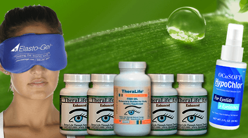
TheraLife
TheraLife has a product line that is oral, all natural when eye drops no longer works. Theralife Eye formula restores normal cellular function from inside out.
To learn more. click here
Management
Because the pathogenesis of meibomian gland dysfunction-associated EDE varies among patients, there is no standard treatment algorithm, and management must be tailored to meet individual needs.2 Initial management should identify and treat the underlying causes of MGD, including rosacea, which can cause blockage of meibomian glands. Once that has been achieved, the treatment approach to MGD should begin with low-risk conservative management before proceeding to higher-risk advanced control. (See “Management of MGD Resulting in EDE” for our suggested general treatment algorithm for the management of patients with MGD.)
Conservative management. These measures include patient education about the condition, behavioral changes such as modifications for or avoidance of exacerbating environments, reduction or elimination of medications that may worsen MGD or EDE, and the use of eyelid hygiene and warm compresses.2,5
Although the recommendation for dietary supplementation with omega-3 fatty acids is first-line therapy for MGD, it may not be beneficial in treating MGD, according to a recent study.6
Over-the-counter eye drops. Topical pharmacological management can target various aspects of the MGD disease process. Non–lipid-containing topical lubricants, such as preservative-free artificial tears, may provide symptomatic relief. However, the duration of their effect is typically limited to a few hours, and the treatment does not address the underlying pathophysiology of MGD.7 Instead, initial treatment should involve replacing the natural oils of the tear film with lipid-containing over-the-counter lubricant eye drops, which may restore the balance of the tear film and provide both symptomatic relief and improvement in clinical signs of MGD and EDE with long-term use.5
Methods to improve meibum flow.
Local, nonpharmacological management consists of mechanically unclogging blocked meibomian glands, thus restoring natural meibum to the ocular surface to prevent fluid evaporation. Four primary methods can do this: warm compresses, thermal pulsation (TPS), intense pulsed light (IPL), and meibomian gland probing (discussed under “Treatment of Refractory Disease“).
Lid hygiene. The most conservative approach to increasing meibum flow is warm compresses, lid scrubs, and gentle massage, performed at home by the patient.3
TPS. Thermal pulsation systems apply heat to the conjunctival surfaces of the upper and lower inner eyelids while putting pulsatile pressure on the outer eyelid surfaces to express the meibomian glands. A study of 20 patients with evidence of meibomian gland obstruction suggested that treatment with TPS has clinically evident benefits up to three years after a single treatment.7
IPL. Several recent studies have demonstrated that IPL, a standard treatment for skin rosacea, also reduces signs and symptoms of EDE in patients with MGD.8
Prescription topical drugs.
Other topical agents for treating MGD include anti-inflammatory medicines (including corticosteroids) and the antibiotic azithromycin.
Azithromycin. Topical azithromycin is helpful for its specific anti-inflammatory effects. One study of 22 patients with symptomatic MGD refractory to eyelid heating and massage demonstrated that topical azithromycin, when applied daily for four weeks, provided symptomatic relief and demonstrated improved signs of eyelid disease in the 17 patients who adhered to the medication regimen.9
THERAPEUTIC APPROACH. This figure lists the various treatment options for MGD-related EDE in the general order of conservative low-risk treatments to advanced higher-risk treatments. The listed treatments may be used individually or in combination. This approach is not proposed as a strict treatment protocol to be followed linearly but rather as a guide to management that should be tailored to the individual patient’s unique pathogenesis of EDE.
Treatment of EDE
Corticosteroids. Brief use of weak topical corticosteroids such as fluorometholone or rimexolone to treat signs and symptoms of the disease that are refractory to other treatments. Although topical corticosteroids do not impact the meibomian glands (typically obstructed but not inflamed in MGD), they reduce the secondary inflammation on the eyelid margins and ocular surface, leading to temporary relief for patients. Patients prescribed topical corticosteroids should be cautioned about the risks of cataract and ocular hypertension or glaucoma with prolonged use. In addition, the physician should limit refills on corticosteroid prescriptions and monitor patients for intraocular pressure elevation.
Systemic medications.
Oral anti-inflammatory drugs and oral antibiotics comprise the systemic pharmacological management options for MGD. Because of their risks, their use should be reserved only for cases of MGD that respond inadequately to the previously mentioned local therapies.
Compared with topical azithromycin, oral doxycycline 100 mg taken twice daily for eight weeks provided relief of MGD symptoms but was not as effective as topical azithromycin in improving signs of gland plugging.10 It has been hypothesized that the mechanism by which these antibiotics improve signs and symptoms of MGD may result from their antibiotic properties reducing bacterial-induced inflammation or of their anticipated effects, which can explain the improvement in lipid content post-treatment.
Intraductal probing. Meibomian gland probing, initially described by Maskin in 2010, helps relieve obstructed meibomian glands by mechanically dilating the glands under local anesthesia.11 In a study of 25 patients, 100% had relief of symptoms four weeks after the procedure. Most patients did not require retreatment during an average follow-up of 11 months. However, this procedure can be painful, so it should be reserved for challenging cases that do not respond to less invasive methods.
Evaporative dry eye
The tear abnormality is created by other periocular diseases, usually leading to increased tear evaporation. Dry eye can also develop when the lacrimal function is normal, and the volume and composition of the lacrimal fluid are sufficient. These conditions are referred to as evaporative dry eye, and each is independently capable of producing dry eye.
Intrinsic causes
Dry eye can be caused by blepharitis, a condition in which contamination by skin lipids causes the tear film to destabilize, increasing tear evaporation. Diseases of the Meibomian gland also increase tear evaporation as insufficient lipids are produced for resurfacing the tear film with each blink. In addition, a qualitative alteration of the Meibomian lipid takes place in such a way as to destabilize the tear film. Dystrophy of the lacrimal glands and a condition called distichiasis (a double row of eyelashes on an eyelid, turned in against the eyeball) can also cause evaporative dry eye.
Lid related disorders
Blinking is essential in maintaining the lipid layer because it forces the lipid from the Meibomian glands and spreads it over the cornea’s surface, sealing in the aqueous layer. Thus, infrequent blinking (for example, in Parkinson’s disease or during excessive use of computer screens) leads to drying of the ocular surface between blinks. Disorders of the lid aperture can also lead to dry eye; for example, the increased width of the palpebral gap that occurs with abnormal protrusion of the eyeball in thyroid disease is associated with ocular drying and tear hyperosmolarity. Lid deformity, resulting in the lid–globe incongruity, leads to poor resurfacing of the ocular tears and is yet another cause of evaporative dry eye.
EXTRINSIC CAUSES
Contact lens wear
Contact lens wear increases tear evaporation from the ocular surface by disrupting the lipid layer of the tear film. This causes irritation, infection, protein deposits, and pain. Research has shown that dry eye is the leading cause of contact lens discomfort.
Surface changes
Any disorder that results in the elevation of the ocular surface is associated with local drying of the cornea. For example, xerophthalmia (where vitamin A deficiency causes the conjunctiva and cornea) is a condition in which tear-deficient and evaporative dry eye can both occur. The disorder is characterized by defective surface wetting associated with abnormal surface changes, including goblet cell loss. The loss of the goblet cells increases dry eye.
Whether tear deficient or tear sufficient, any of the disorders discussed so far may occur in conjunction with any other – and several of them commonly do. For example, lacrimal gland deficiency may be accompanied by Meibomian gland deficiency. At the same time, cicatricial conjunctival disease may cause dry eye by occlusion of the lacrimal gland ducts and causing a lid incongruity that interferes with tear resurfacing with each blink. When the signs of more than one form of dry eye are present, it may not always be possible to differentiate their relative contribution to the dry eye condition.
Distinguishing Evaporative from Aqueous Deficient Dry Eye
The accurate diagnosis of dry eye syndrome (DES) and the classification of its severity and subtype are essential to developing an effective treatment plan. DES has been diagnosed more readily today thanks to increasing awareness of the syndrome and its many irritating symptoms and a plethora of diagnostic screening methods. DES can significantly affect our patients’ vision and quality of life. It should be detected and treated as early as possible to avoid other long-term side effects.
The initial diagnosis and management of DES involve determining the presence of the condition; only then can the presenting subtype, either aqueous-deficient dry eye (ADDE) or evaporative dry eye (EDE), be identified. These subtypes can be present individually or in combination, but unfortunately, some objective tests are specific to only one subtype, such as the Schirmer test, tear turnover, and phenol red for ADDE or lid margin examination for EDE. Other tests simply are for manifestations that appear only later in the disease process, such as corneal and conjunctival staining.
Corneal and conjunctival staining. In patients with early or mild dry eye, standard objective tests often fail to diagnose DES; however, the accuracy of diagnosis improves with increasing severity of DES. In addition to determining which DES subtype is present, it is important to assess the severity to initiate an optimal treatment plan.
CHARACTERISTICS
Both ADDE and EDE give rise to similar signs, leading to increased evaporation and reduced tear film stability. But distinguishing between these subtypes and whether they exist individually or in combination is crucial. EDE is the most common type of dry eye, accounting for 35% to 45% of cases,1-4, but both forms of dry eye coincide in the most severe cases.
ADDE. The cause of ADDE is a lack of aqueous tear secretion by the lacrimal glands. This can result in a concentrated tear film (hyperosmolarity) and an unstable tear film with desiccation of the ocular surface.
EDE. This dry eye subtype is most often caused by meibomian gland dysfunction (MGD). The lipid secretion required to control evaporation and maintain a normal tear film is abnormal. EDE can cause excessive evaporative tear loss at an early stage of the disease with increased tear osmolarity and tear film instability and thus present with signs similar to ADDE.5
In contrast to the DES seen in most patients, certain forms present with different characteristics, including those associated with autoimmune diseases. In Sjögren syndrome, autoantibodies target exocrine glands such as the lacrimal glands, affecting tear production. Sjögren syndrome can also occur secondary to systemic autoimmune diseases like rheumatoid arthritis or lupus. Graft-versus-host disease (GVHD) is another systemic autoimmune disease that can go undetected if not screened for and treated accordingly. These autoimmune diseases, all distinguished by a severe inflammatory component during at least a set interval of the disease course, are associated with ADDE and EDE.
DIAGNOSE ACCURATELY
Our priority as physicians is to determine if DES is present. Once the diagnosis is made, we can then decide which subtypes are present and at what severity. Symptoms often point toward one form (ADDE or EDE). Still, symptoms alone can be inadequate for a differential diagnosis of DES. The symptoms characteristic of dry eye can be experienced with a range of ocular surface conditions and tear film disorders. They are subject to psychological overlays and influenced by alterations in sensory receptors in DES. When DES is suspected, an evaluation of the eyelids, the tear film, the ocular surface, and the lacrimal glands should be performed. This is the best way to determine the etiology.
DES is a condition with various risk factors, including age, androgen deficiency, chronic environmental stress, changes in blinking patterns, systemic autoimmune disease, the use of systemic drugs, nerve damage caused by surgery, and contact lens wear leading to instability and tear hyperosmolarity.5 According to the Dry Eye WorkShop, tear hyperosmolarity in ADDE results from reduced tear production from the lacrimal function unit with a normal evaporation rate. With EDE, in contrast, increased evaporation occurs. Increased tear osmolarity is the principal feature causing damage to the epithelial surface and triggering a cascade of inflammatory events, including an increase in inflammatory tear cytokines and apoptotic cell death. It is also responsible for initiating alterations in mucin expression, leading to ocular surface damage.6
PREFERRED SCREENING METHODS
Physicians have difficulty diagnosing the early stages of DES because onset and progression are typically gradual. Existing tests evaluate various characteristics of the tear film. Still, physicians should use both experience and an adequate testing method to distinguish between the two forms of DES.
Conventional screening. For ADDE and EDE, traditional objective testing such as the Schirmer test, tear break-up time (TBUT), noninvasive tear break-up time (BUT), and ocular surface staining produce variable results due to instability of the tear film in patients with dry eye. On the other hand, Tear osmolarity is a global test for the presence of DES because it measures the concentration of the tear film, which is high in all forms of DES. One tear osmolarity test is the TearLab Osmolarity System (TearLab Corp., San Diego). Although it does not differentiate between ADDE and EDE, it is an excellent measure of disease severity and response to treatment.
Tests to diagnose EDE and MGD. Tests for EDE can also diagnose MGD. These include grading lid margin disease for vascularity and lipid expression using the expression test described by Korb and colleagues. In the diagnostic expression test, pressure is applied to the outside of the eyelid, similar to the amount of pressure the lid muscles exert during the blink motion (1.25g/mm2). Pressure is applied manually with a finger against any kind of suitable instrument (e.g., gland expressor) placed on the conjunctival side of the lid.1
Noninvasive tests. Another test, the LipiView interferometer (TearScience Inc., Morrisville, North Carolina), objectively quantifies lipid layer thickness by capturing images of the tear film. This noninvasive test, which uses an automated measure of a tear sample’s interferometric colors to indicate the thickness of the tear film’s lipid layer, can be used to identify lipid abnormalities, which are pathognomonic for MGD.
Patient questionnaires. Surveys can quantify the frequency and severity of patients’ symptoms. For instance, the Standard Patient Evaluation of Eye Dryness (SPEED) questionnaire provides a means for patients to explain their symptoms, allowing both patient and physician to establish insight into the patient’s complaints and how these symptoms affect their quality of life.
AVAILABLE TREATMENTS
Management of DES varies from patient to patient and according to the severity of symptoms and ocular signs. Specific treatments are available to address each subtype of DES. For ADDE, artificial tear solutions lubricate the eye, stabilize the tear film, and retard evaporative tear loss. Punctal plugs and anti-inflammatory and immune-modulating drugs are also used primarily to treat ADDE. Lid scrubs and external heat are mainly used to treat EDE and MGD.
The LipiFlow System (TearScience Inc.) is another treatment for EDE. For more information, see Thermal Pulsation to Relieve Meibomian Gland Blockages.
Evaporative dry eye
Dry eye syndrome is an uncomfortable condition caused by a lack of quality tears. Evaporative dry eye (EDE) is the most common form of dry eye syndrome. It’s usually caused by a blockage of the oil glands that line the margins of your eyelids. These tiny glands, called meibomian glands, release oil to cover your eye surface and prevent your tears from drying out.
Keep reading to learn more about EDE.
Conclusions
Refined diagnostic and clinical accuracy is essential in treating dry eye. Does evaporation cause dry eyes? Since ADDE alone is thought to be a vital contributor to EDE, some drugs viewed by others as more useful for EDE may prove helpful in treating ADDE.