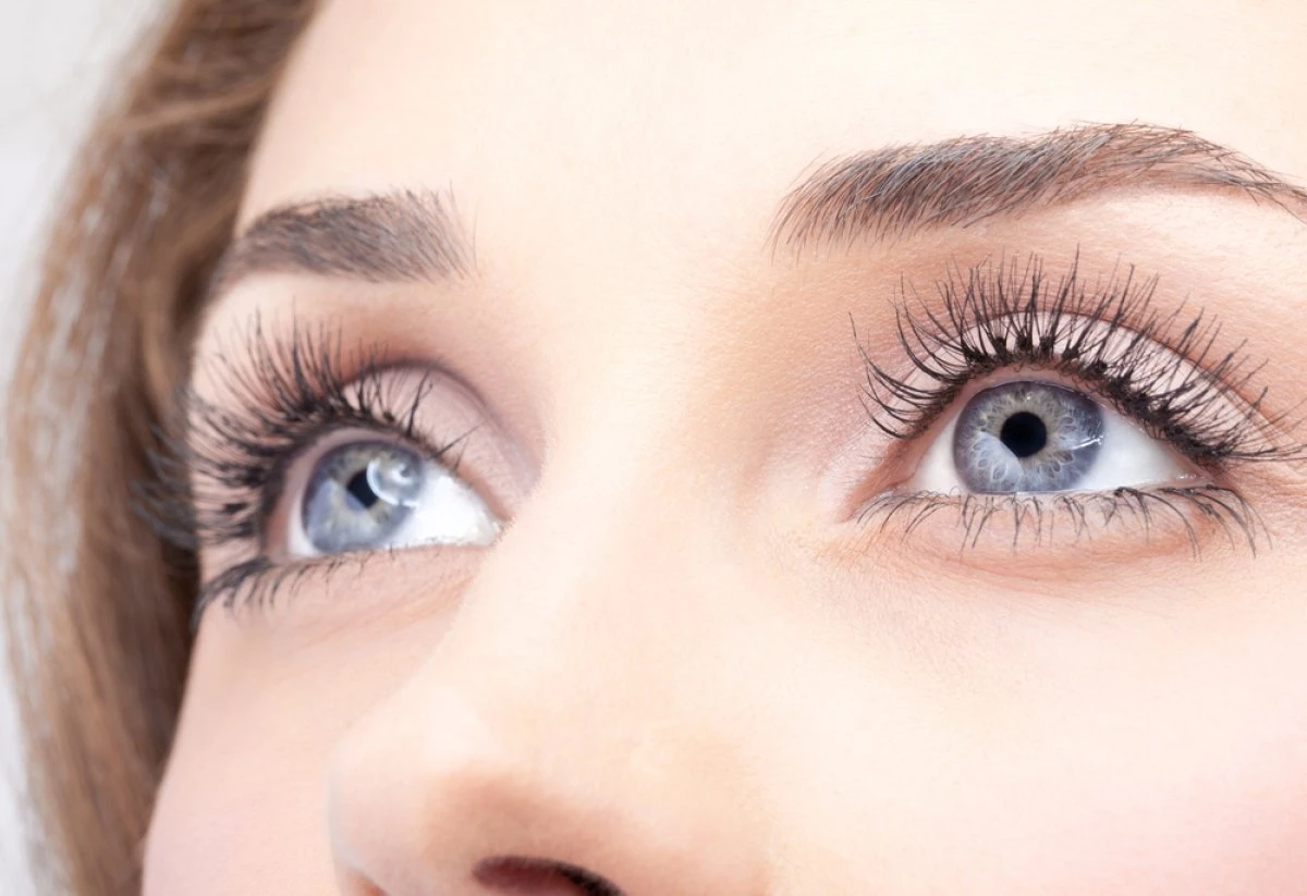
Are you experiencing dry, irritated eyes? Eyes may be light-sensitive, often blurry vision, and eyes may even be watery? Are you over 50 and maybe in menopause? Have you had LASIK surgery, or wearing contact lenses?
The chances are pretty good that you have chronic dry eyes.
Causes
There are many reasons why dry eyes develop, including:
- Age – As a person ages naturally, you will experience some of the symptoms of dry eyes
- Gender – Due to hormonal changes during pregnancy, menopause, or the use of oral contraceptives, dry eyes often happen in women.
- Medication –Medications, such as antihistamines, antidepressants, and blood pressure medications can cause a dry eye to reduce tear production.
- Medical Conditions – Certain medical problems such as arthritis, diabetes, Sjogren’s, and thyroid issues can lead to dry eyes.
- Environment –Windy, bright sunlight, dry air, or pollutants can increase the development of dry eyes.
- Computers – Long hours of using computers without frequent blinking can lead to dry eyes
- Corrective Measures – LASIK surgery or wearing contact lenses can cause dry eyes.
- Abnormal Eye Conditions – Inflammation of the eyelids – blepharitis, Meibomian Gland Dysfunction, or a defect of the eyelids can all lead to dry eyes.
Symptoms
These dry eye symptoms can affect one or both eyes and include:
- Scratchy, stinging, burning sensation and eye pain in one or both eyes
- Thick, stringy mucus around or in the eyes
- Sensitivity to light- Photophobia
- Red eyes
- Foreign body sensation- feels like something is in the eye; actually, there is none.
- Contact lenses are getting dirty often, and eyes are feeling dry.
- Difficulties driving at night.
- Watery eyes
- Blurred vision
- Tired irritated eyes
If any of these symptoms persist, see an eye doctor for the correct diagnosis. Visit TheraLife for a complete line of products that help relieve and even eliminate these symptoms.
There is no cure for dry eyes, but we can relieve the symptoms and put your life back on track.
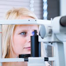
Diagnosis
A series of comprehensive eye examinations by a qualified eye doctor can test for the quality and quantity of tear production. Some tests determine health problems, medications, or outside factors that may contribute to dry eyes.
Examples of the tests are:
- Schirmer’s Test – Using a phenolphthalein dye thread under the eyelid to determine tear production determines dry eye syndrome. If the eyes produce too many or too few tears, this test can help determine the best treatment options. Normal tear production is 15-20mm of tear in 10 minutes.
- Rose Bangel Stains – to determine the damage to epithelial cells with this stain placed on the eye surface. Rose Bangel stains aid in the diagnosis of preocular tear film disorders, mucin preocular film deficiencies. Place Stain eye drops in the eyes for 3 to 5 minutes. Stain patterns can determine the damage to the epithelial cells.
- Tear Break-up Test – Measures the viscosity of tear. How thick the tear is while coating the eyeball. Fluorescein is applied, and the time it takes the dye to disperse is measured. Other methods to measure tear viscosity include Keratometer, Keratoscope, or Telescope.
Once the causes are determined, the eye doctor can prescribe proper treatment plans.
TheraLife has many different products available to treat a wide variety of dry eye symptoms and causes.
Treatment with TheraLife
Frequent use of eye drops makes eyes drier. Although over-the-counter eye drops can provide temporarily relieve, those who suffer from chronic dry eyes need a long-term solution. Therefore, TheraLife’s approach is oral treatment. We are treating dry eyes from the inside out.
As a chronic dry eye relief leader, TheraLife addresses the root cause instead of masking the symptoms. The root cause is underactive tear production and inflammation. TheraLife provides complete relief by helping the body produce its own tears, eliminating inflammation and dryness.
It is clinically proven to work for 80% of first-time users. The proprietary TheraLife Eye formula restores and revitalizes tear secretion from a cellular level to allow the eyes to produce balanced and sustainable tears. Stop suffering from constant dry, red, and irritated eyes.
Let TheraLife put you on the right path to complete relief.
Customer Story
Chronic Severe Dry Eyes – Relief by TheraLife
“Your TheraLife Eye products are the answers to my prayers for relief of the chronic dry eyes I have suffered for so long. I have tried every lubricating eye drops, punctal plugs, Restasis, etc., with no help. What made it worse was some of the conventional eye doctors told me there is no cure for dry eyes. This statement gave me more challenge to seek another alternative way of healing, and I thank God that I found your website. You have saved my eyes, my self-esteem, and my self-worth.
I cannot thank you enough for these fantastic products you have formulated—my heartfelt thanks and gratitude from the bottom of my heart”
E.B. Clarksville, TN
*Results may vary
For more customer stories, click here
References
- Stapleton F, Alves M, Bunya VY, et al. : TFOS DEWS II Epidemiology Report.Ocul Surf. 2017;15(3):334–65. 10.1016/j.jtos.2017.05.003 [
- Blehm C, Vishnu S, Khattak A, et al. : Computer vision syndrome: a review.Surv Ophthalmol. 2005;50(3):253–62. 10.1016/j.survophthal.2005.02.008
- Tsubota K, Nakamori K: Dry eyes and video display terminals.N Engl J Med. 1993;328(8):584. 10.1056/NEJM199302253280817
- Uchino M, Dogru M, Uchino Y, et al. : Japan Ministry of Health study on prevalence of dry eye disease among Japanese high school students.Am J Ophthalmol. 2008;146(6):925–9.e2. 10.1016/j.ajo.2008.06.030
- Zhang Y, Chen H, Wu X: Prevalence and risk factors associated with dry eye syndrome among senior high school students in a county of Shandong Province, China.Ophthalmic Epidemiol. 2012;19(4):226–30. 10.3109/09286586.2012.670742
- Uchino M, Yokoi N, Uchino Y, et al. : Prevalence of dry eye disease and its risk factors in visual display terminal users: the Osaka study.Am J Ophthalmol. 2013;156(4):759–66. 10.1016/j.ajo.2013.05.040
- Uchino M, Uchino Y, Dogru M, et al. : Dry eye disease and work productivity loss in visual display users: the Osaka study.Am J Ophthalmol. 2014;157(2):294–300. 10.1016/j.ajo.2013.10.014
- Asiedu K, Dzasimatu SK, Kyei S: Impact of Dry Eye on Psychosomatic Symptoms and Quality of Life in a Healthy Youthful Clinical Sample.Eye Contact Lens. 2018;44 Suppl 2:S404–S409. 10.1097/ICL.0000000000000550
- Jonas JB, Wei WB, Xu L, et al. : Self-rated depression and eye diseases: The Beijing Eye Study.PLoS One. 2018;13(8):e0202132. 10.1371/journal.pone.0202132
- Galor A, Felix ER, Feuer W, et al. : Dry eye symptoms align more closely to non-ocular conditions than to tear film parameters.Br J Ophthalmol. 2015;99(8):1126–9. 10.1136/bjophthalmol-2014-306481
- Kawashima M, Uchino M, Yokoi N, et al. : Associations between subjective happiness and dry eye disease: a new perspective from the Osaka study.PLoS One. 2015;10(4):e0123299. 10.1371/journal.pone.0123299
- Um SB, Yeom H, Kim NH, et al. : Association between dry eye symptoms and suicidal ideation in a Korean adult population.PLoS One. 2018;13(6):e0199131. 10.1371/journal.pone.0199131
- Schiffman RM, Walt JG, Jacobsen G, et al. : Utility assessment among patients with dry eye disease.Ophthalmology. 2003;110(7):1412–9. 10.1016/S0161-6420(03)00462-7
- McDonald M, Patel DA, Keith MS, et al. : Economic and Humanistic Burden of Dry Eye Disease in Europe, North America, and Asia: A Systematic Literature Review.Ocul Surf. 2016;14(2):144–67. 10.1016/j.jtos.2015.11.002
- Yu J, Asche CV, Fairchild CJ: The economic burden of dry eye disease in the United States: a decision tree analysis.Cornea. 2011;30(4):379–87. 10.1097/ICO.0b013e3181f7f363
- Lemp MA: Report of the National Eye Institute/Industry workshop on Clinical Trials in Dry Eyes.CLAO J. 1995;21(4):221–32.
- Craig JP, Nichols KK, Akpek EK, et al. : TFOS DEWS II Definition and Classification Report.Ocul Surf. 2017;15(3):276–83. 10.1016/j.jtos.2017.05.008
- Luo L, Li DQ, Corrales RM, et al. : Hyperosmolar saline is a proinflammatory stress on the mouse ocular surface.Eye Contact Lens. 2005;31(5):186–93. 10.1097/01.ICL.0000162759.79740.46
- Lanza NL, Valenzuela F, Perez VL, et al. : The Matrix Metalloproteinase 9 Point-of-Care Test in Dry Eye.Ocul Surf. 2016;14(2):189–95. 10.1016/j.jtos.2015.10.004
- Yoon KC, Jeong IY, Park YG, et al. : Interleukin-6 and tumor necrosis factor-alpha levels in tears of patients with dry eye syndrome.Cornea. 2007;26(4):431–7. 10.1097/ICO.0b013e31803dcda2
- Niederkorn JY, Stern ME, Pflugfelder SC, et al. : Desiccating stress induces T cell-mediated Sjögren’s Syndrome-like lacrimal keratoconjunctivitis.J Immunol. 2006;176(7):3950–7. 10.4049/jimmunol.176.7.3950
- McClellan AJ, Volpe EA, Zhang X, et al. : Ocular surface disease and dacryoadenitis in aging C57BL/6 mice.Am J Pathol. 2014;184(3):631–43. 10.1016/j.ajpath.2013.11.019
- Galor A, Feuer W, Lee DJ, et al. : Ocular surface parameters in older male veterans.Invest Ophthalmol Vis Sci. 2013;54(2):1426–33. 10.1167/iovs.12-
- Gipson IK: Distribution of mucins at the ocular surface.Exp Eye Res. 2004;78(3):379–88. 10.1016/S0014-4835(03)00204-5
- Gipson IK, Hori Y, Argüeso P: Character of ocular surface mucins and their alteration in dry eye disease.Ocul Surf. 2004;2(2):131–48. 10.1016/S1542-0124(12)70149-0
- Cher I: A new look at lubrication of the ocular surface: fluid mechanics behind the blinking eyelids.Ocul Surf. 2008;6(2):79–86. 10.1016/S1542-0124(12)70271-9
- Willcox MDP, Argüeso P, Georgiev GA, et al. : TFOS DEWS II Tear Film Report.Ocul Surf. 2017;15(3):366–403. 10.1016/j.jtos.2017.03.
- Willshire C, Buckley RJ, Bron AJ: Central Connections of the Lacrimal Functional Unit.Cornea. 2017;36(8):898–907. 10.1097/ICO.0000000000001250
- Cox SM, Nichols JJ: The neurobiology of the meibomian glands.Ocul Surf. 2014;12(3):167–77. 10.1016/j.jtos.2014.01.005
- Mantopoulos D, Cruzat A, Hamrah P: In vivoimaging of corneal inflammation: new tools for clinical practice and research. Semin Ophthalmol. 2010;25(5–6):178–85. 10.3109/08820538.2010.518542
- Knop E, Knop N, Millar T, et al. : The international workshop on meibomian gland dysfunction: report of the subcommittee on anatomy, physiology, and pathophysiology of the meibomian gland.Invest Ophthalmol Vis Sci. 2011;52(4):1938–78. 10.1167/iovs.10-6997c
- Baudouin C, Messmer EM, Aragona P, et al. : Revisiting the vicious circle of dry eye disease: a focus on the pathophysiology of meibomian gland dysfunction.Br J Ophthalmol. 2016;100(3):300–6. 10.1136/bjophthalmol-2015-307415
- Jester JV, Nicolaides N, Kiss-Palvolgyi I, et al. : Meibomian gland dysfunction. II. The role of keratinization in a rabbit model of MGD.Invest Ophthalmol Vis Sci. 1989;30(5):936–45.
- Rajagopalan K, Abetz L, Mertzanis P, et al. : Comparing the discriminative validity of two generic and one disease-specific health-related quality of life measures in a sample of patients with dry eye.Value Health. 2005;8(2):168–174. 10.1111/j.1524-4733.2005.03074.x
- Hwang HS, Parfitt GJ, Brown DJ, et al. : Meibocyte differentiation and renewal: Insights into novel mechanisms of meibomian gland dysfunction (MGD).Exp Eye Res. 2017;163:37–45. 10.1016/j.exer.2017.02.
- Jester JV, Brown DJ: Wakayama Symposium: Peroxisome proliferator-activated receptor-gamma (PPARγ) and meibomian gland dysfunction.Ocul Surf. 2012;10(4):224–229. 10.1016/j.jtos.2012.07.001
- Sullivan DA, Sullivan BD, Evans JE, et al. : Androgen deficiency, Meibomian gland dysfunction, and evaporative dry eye.Ann N Y Acad Sci. 2002;966:211–22. 10.1111/j.1749-6632.2002.tb04217.x
- Bron AJ, de Paiva CS, Chauhan SK, et al. : TFOS DEWS II pathophysiology report.Ocul Surf. 2017;15(3):438–510. 10.1016/j.jtos.2017.05.011
- Bozkurt B, Irkeç MT, Atakan N, et al. : Lacrimal function and ocular complications in patients treated with systemic isotretinoin.Eur J Ophthalmol. 2002;12(3):173–6.
- Chhadva P, Goldhardt R, Galor A: Meibomian Gland Disease: The Role of Gland Dysfunction in Dry Eye Disease.Ophthalmology. 2017;124(11S):S20–S26. 10.1016/j.ophtha.2017.05.031
- Belmonte C, Nichols JJ, Cox SM, et al. : TFOS DEWS II pain and sensation report.Ocul Surf. 2017;15(3):404–37. 10.1016/j.jtos.2017.05.002
- Galor A, Covington D, Levitt AE, et al. : Neuropathic Ocular Pain due to Dry Eye is Associated with Multiple Comorbid Chronic Pain Syndromes.J Pain. 2016;17(3):310–8. 10.1016/j.jpain.2015.10.019
- Lee CJ, Levitt RC, Felix ER, et al. : Evidence that dry eye is a comorbid pain condition in a U.S. veteran population.Pain Rep. 2017;2(6):e629. 10.1097/PR9.0000000000000629
- Aggarwal S, Kheirkhah A, Cavalcanti BM, et al. : Autologous Serum Tears for Treatment of Photoallodynia in Patients with Corneal Neuropathy: Efficacy and Evaluation with In VivoConfocal Microscopy. Ocul Surf. 2015;13(3):250–62. 10.1016/j.jtos.2015.01.005
- Sivanesan E, Levitt RC, Sarantopoulos CD, et al. : Noninvasive Electrical Stimulation for the Treatment of Chronic Ocular Pain and Photophobia.Neuromodulation. 2017. 10.1111/ner.12742
- Diel RJ, Kroeger ZA, Levitt RC, et al. : Botulinum Toxin A for the Treatment of Photophobia and Dry Eye.Ophthalmology. 2018;125(1):139–40. 10.1016/j.ophtha.2017.09.031