Evaporative dry eye is a dry-eye condition that causes swollen eyelids and blurred vision.
This usually happens because the eye is not releasing sufficient tears to cover the ocular surface adequately.
Evaporative dry eye is quite common accounting for over 90% of true sources of dry eye syndrome.
Sometimes the evaporative dry eye aqueous deficit causes an eye that produces no tears.
Introduction- Evaporative dry eye
Dry eye disease is widespread and affects quality of life.
There are several underlying causes of ocular surface diseases including loss of homeostasis and ocular inflammation that occur on the tear film and ocular surface inflammation and damages.
It’s an overwhelming burden for healthcare systems as the leading reason for patient visits.
Summary- Evaporative dry eye
Evaporative dry eye disease affects a fifth of the world’s population with adverse health effects.
Most people have mild disease.
This disorder is multifactorial with a significant inflammatory component causing significant damage to vascular and ocular surface tissues.
Meibomian gland dysfunction can become invasive in dry eye and may be the cause of inflammation of ophthalmologic tissues.
Causes of Inadequate Tears
These two main etiologies of DED are known as aqueous deficiency dry eye and evaporative dry eye (EDE), respectively.
EDE
The pathophysiology of EDE includes lid-related causes such as meibomian gland dysfunction (MGD) and blink problems, as well as ocular surface–related causes including mucin and contact lens dysfunction.
By far, the principal cause of dry eye is MGD.
A growing problem that is now accounts for up to two-thirds of all cases of dry eye.
EYE WITH MGD.
Strings of waxy meibum exuded from dysfunctional meibomian glands. MGD The meibomian glands.
Eyelids contain a number of small glands – called meibomian glands located on the upper and lower eyelids – which provide oil to the tear film. The glands have an opening in the margins of the eyelids that lies inside it.
Depending on what the condition is, the gland can be clogging or not performing properly.
Meibomian gland dysfunction are common.
Mild cases are sometimes undiagnosed and do not have the proper treatment.
Evaporative
Excessive dryness in eyes occurs when the tear tissue lacks a lipid layer, increasing tear evaporation.
Usually this occurs in more than 85 per cent of dry eye diseases.
Blepharitis, a lid margin inflammation, causes a meibomian gland malfunction.
Blepharitis is a rare disorder characterized by rosacea in eosinophilic eyes and atopy, seborrhaeic dermatitis, staphylococcus infections and depression with mites.
Tear depletion affects the immune system and causes inflammation, so dry eye diseases are both causes and effects of Blepharitis.
The Tear Film
EVAPORATIVE DRY EYE occurs due to tear film instability, and evaporates faster than normal (less than 10 seconds).
To understand evaporative dry eye, you need to know a little about what your tears are actually made of.
The tear film is thought to be made up of a mucin or mucus layer that coats the surface of the cornea and makes tears “stick” to the eye.
The next layer is composed of water and oil. Oil helps lubricate the eye balls.
How does evaporation dry eye work?
A mucin film on eyelashes may contain mucus and mucous that coat corneal surface making tears splinter on eyelashes.
The next layer comprises water and oil. Oil prevents the formation of tear films.
Upon opening your eyes during waking time tears evaporate in a drainage system.
When you have an open eye you experience a lot of evaporated liquid.
If you lose the oils from your tear films the tears will evaporate quickly.
The Tear Film and Ocular Surface Society (TFOS) defines DED as “a multifactorial disease of the ocular surface characterized by a loss of homeostasis of the tear film and accompanied by ocular symptoms, in which tear film instability and hyperosmolarity, ocular surface inflammation and damage and neurosensory abnormalities play etiological roles.
Dry eye disease occurs in two main categories: aqueous deficiencies and the evaporative form (e.g. Both can coexist) .
Your eye doctor will perform Diagnostic Tests to Evaluate Ocular Surface.
Symptoms of Evaporative dry eye
Symptoms include: fluctuating and unstable vision, redness, irritation, “tired eyes”, pain, or discomfort .
Eye pain, burning, or redness Itchy eyes Feeling like there’s sand or dirt in your eyes Blurry vision Eyes that feel tired after reading Trouble wearing contact lenses Not being able to produce tears when you cry
Risk factors of Evaporative dry eye
A variety of factors can cause dry eye disease.
Female gender had the highest prevalence of all surveyed risk factors, and the prevalence for women nearly doubled for women and the prevalence for both genders.
The aging process may have been caused mainly by reduced androgen levels, because androgen upregulated meibomian gland function in animals.
The effect of estrogen on the skin is unknown but is likely to reduce dryness.
About 10% of people with dry eye have Sjogren’s. This syndrome is associated with rheumatoid arthritis, an autoimmune disorder.
Other health problems that can cause aqueous tear-deficient dry eye include: Viruses Hepatitis C HIV and AIDS Lymphoma Sarcoidosis Hemochromatosis Amyloidosis Graft versus host disease Damage to tear glands or tear ducts
Meibomian gland dysfunction
Myibomian glands located at the upper eyelids secrete a protective fat layer that helps prevent evaporation from aqueous tears.
The glands can be blocked and cause eye irritations and dry eye as the result of the eroding of aqueous tears. This is called meibomian gland dysfunction.
Diagnosis and assessment
Identifying dry eye disease with precise accuracy can be complicated if the presentation is variable.
Patients often report non-specific symptoms, which include irritable eyes and skin sensation, irritation and burning.
It is possible that excessive watery sensation is due to discomfort triggered by reflex tearing. It can not be easily determined whether symptoms have a high intensity in the eyes or whether there are symptom-depleted corneal acuities.
Diagnosis of Evaporative dry eye
Evaporative dry eye can be diagnosed with slitlamp bio-microscopes. At high magnification, doctors are also allowed to see individual openings in meibomian glands.
Occasionally the gland is filled with dirt.
When myebomian gland dysfunctions are chronic they may even become atrophy. The consistency and number of tears are also analyzed.
Unless dry eye is evaporated tears may appear thin or frothy.
Tear osmolarity is the single best metric to diagnose and classify DED. A positive result is any reading greater than 308mOsm/L or a difference of more than eight between eyes.
Although the pathophysiology of patients with EDE or ADDE differs, the tear osmolarity and distribution do not.
Ocular surface staining . Conjunctival and lid margin damage is best viewed with lissamine green staining, and corneal damage is more visible with fluorescein dye.
Rose bengal concentrates on corneal and conjunctival cells that lack mucin and can identify damaged epithelial cells.
Staining in either eye confirms a dry eye disease
Clinical assessment and various tests are required to diagnose dry eye disease.
Ocular surface staining In dry eye disease, there is loss of the protective glycocalyx barrier, due to increased shedding of the corneal and conjunctival epithelial cells.
The new underlying cells are able to absorb vital dyes, with the degree of pathological staining matching disease severity.
Punctal Plugs
Commonly, punctal occlusion is performed via punctal plugs.
Absorbable, collagen-based plugs with absorption rates that vary from one week to six months are the most commonly used.
Non-absorbable, or “permanent,” plugs are typically silicone-based.
For advanced cases, permanent surgical closure of the puncta via total or partial thermal cauterization, occlusion with a conjunctival flap, punctal suturing or total destruction of the canaliculus can be performed.
Punctal occlusion is most successful when combined with other DED treatments
Treatment of Refractory Disease (LASIK)
Corticosteroid. A brief treatment with weakened topical corticosteroid like fluorometholone can be used in patients with refractory conditions.
Although topical corticosteroid doesn’t affect myoma itself, they reduce secondary inflammation around the ophthalmic surfaces and eyelash margins, which causes temporary relief.
Topical corticosteroids can increase glaucoma and cataract if they are used for a long duration.
After LASIK, you may experience decreased corneal sensation, which can be triggered pharmacologically, after refractive surgery or secondary to disease.
Treatment of Evaporative dry eye
Depending on the patient’s symptoms, he can take several different treatments including medication or surgical treatments for dry eye.
Artificial tears-
These eye drops can be purchased over the counter.
Artificial Tears / Liquid Gel / Ointment: use up to 4x/day. If greater than 4x/day, PRESERVATIVE FREE eye drops are recommended.
For a cool and soothing effect when the eye drops are placed on the eyes, store your tears in the refrigerator.
In more severe disease, the dilution effect is attenuated due to reduced tear volume, so preservative-free eye drops are recommended.
The Tear Film
To understand evaporative dry eye, you need to know a little about what your tears are actually made of.
The tear film is thought to be made up of a mucin or mucus layer that coats the surface of the cornea and makes tears “stick” to the eye.
The next layer is composed of water and oil. Oil helps prevent tears from evaporating.
Omega 3 fatty acids
A NIH study recently found Fish Oil alone (Omega 3 fatty acids) to be ineffective: https://www.nih.gov/news-events/news-releases/omega-3s-fish-oil-supplements-no-better-placebo-dry-eye.
Omega 3 is effective when used in combination with other dry eye treatments, like TheraLife.
Warm compresses
About Warm Compresses – it heats up to soften the clogs in the oil glands for easy removal.
Autologous serum drops
In more advanced cases of DED where topical pharmaceutical agents do not provide adequate relief of severe ocular surface disease, autologous serum is an effective option.
It is derived from the biomarkers in a patient’s own blood and contains many biochemical characteristics similar to that of human tears, such as pH level, nutrients, vitamins, albumin, fibronectin and epithelial stimulating factors.
Intranasal tear neurostimulator
Intranasal tear neurostimulation induces normal tear production by stimulating the nasolacrimal reflex.
The nasolacrimal reflex up-regulates tear production following chemical or mechanical stimulation of the nasal mucosa.
The TrueTear Intranasal Tear Neurostimulator (Allergan) temporarily increases tear production during neurostimulation in adults.
This device consists of a handheld stimulator unit equipped with a disposable two-pronged hydrogel.
Management
The quantity and quality of tears produced are monitored to verify if the patient’s tear production is operating under the acceptable normal level.
Quick remedies suggested to cure evaporative dry eye problem if the meibomian glands are not performing well, the person who has acquired a mild case of this type of eye disorder is suggested to do warm compresses as well as eyelid hygiene with special lid cleansing pads to improve gland performance.
Because pathogenesis of MGD-associated EDE differs between individuals, there is no standard treatment algorithm, and treatment should suit individual needs.
In initial management MGD may cause rosacea, which may block meibomian gland function.
After that, treatment of MGD must start with low-risk conservative management, then move onto high-risk advanced management.
The general treatments algorithms are suggested for MGD treatment. Conservative management.
Home remedies
Take supplements.
You might try fish oil or flaxseed oil, which are rich in omega-3 fatty acids. Before you take any pills, get your doctor’s OK and ask what dosage is right for you.
Avoiding being around smoke:
Cigarette smoke and other fumes can irritate the eyes and eyelids and reduce tear production. This can cause the oils to back up and clog in the glands.
When you’re inside, stay out of the breeze created by ceiling fans or portable fans.
Be aware of how often you blink. Try to do it more, especially if you’re looking at a computer screen. It’s easy to forget.
Don’t smoke. Steer clear of smoky rooms and people who smoke. Show Sources SOURCES: The Ocular Surface : “The Definition and Classification of Dry Eye Disease: Report of the Definition and Classification Subcommittee of the International Dry Eye Work Shop (2007).
Conclusion
MGD management is multi-faceted and should be tailored to meet patient requirements.
Several of the described treatment methods have proven effective on the edema mediated by the MGH molecule.
Clinicians will sometimes recommend several therapies for different symptoms. 1 Javadis MA & Feizi. A review of the J Ophthalmology Res 11(3) 190-193. 3. Craig, PJ, and colleagues. Optik surfs. 2016;17(4):808-812. 2. “Finale”. ocul surf. 2012: 142-154. 4. Nelson MJ & Co. The study of eye health is done with a microscope in ophthalmology and in vascular sciences. 2012;52(4): 1931-39. 4. Geerling J and others. Invest ocular sct Sciol. 2012 54.2: 2084-5241. I am an entrepreneur. Asbell P.A. & Co. J Physiol Med J. 2018; 3381818:16481-1690. 8. Greiner JJ.
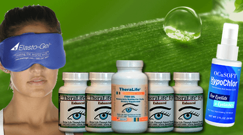
Theralife Eye Treatment for chronic dry eyes.
All natural oral treatment that works.

Fast relief for dry eyes with TheraLIfe