Powerful Treatment for Clogged Meibomian Glands with TheraLife
Meibomian oil glands (Meibomian Gland Dysfunction – MGD) become clogged due to chronic dry eyes and blepharitis.
TheraLife treats dry eyes, blepharitis and MGD simultaneously to achieve optimum results. No more drops

Customer Stories
Blepharitis/Meibomian Gland Dysfunction – Relief in 2 weeks.
Two weeks since I started the Theralife Enhanced and I must say that I have experience everything you mentioned in your email:
- Moist eye
- less sensitivity to light
- less grittyness in my eye
I will continue taking the recommended number of tablets each day. The last thing I want to do is to start over…
I went to see my doctor yesterday. He said I have ocular rosacea. He prescribed the AzaSite eye drop and doxcycline Monohydrate oral. From what I read, AzaSite is very effective for treating MGD.
I cant wait for the day when I don’t have to think about my eyes all the time…
I will keep you informed for my progress.
Thank you
D.A Kenmore, WA USA
Introduction
This manual method of unclogging the meibomian gland is called “Debridement – Scaling.”
Clogged meibomian glands are a significant problem in people with chronic dry eye disease. The meibomian glands get inflamed and clog up the pores to prevent lubricants from flowing.
Lid Margin Debridement – Scraping and cleaning meibomian glands is a new simple procedure. It uses a lid paddle to gently scrape along the eyelids to remove the dead epithelial cells and open up the meibomian glands. You can see the fluid flow out immediately.
At one month post-treatment, people reported a significant improvement in dry eye symptoms.
The researchers showed there is an increase in the number of functional meibomian glands. debridement is a promising development in Meibomian Gland Dysfunction (MGD) therapy\
How do meibomian glands get clogged?
MGD becomes more common as we age. In cases of moderate to advanced Meibomian Gland Dysfunction, devitalized epithelial cells can accumulate on the lid margin over the meibomian gland orifices. Clogging of orifices is similar to dead skin cells blocking facial pores. Significant accrual of this material can create an almost plaque-like quality to the lid margin that will stain with lissamine green. There should be no surprise that meibomian gland function is sub-optimal in this environment. Eyelid hygiene does very little to address it.
A painless, quick, in-office procedure can remove this debris, which allows for improved gland function and better response to warm compress therapy. The eye is numbed with a drop of topical anesthetic, and the lid margins are ‘exfoliated’ with a spud by gently scraping across the length of the margin. You can often observe glands immediately release their contents as you pass over them, even when months of warm compresses have failed to do the same.
Debridement can treat anterior blepharitis of the lid margin.
How does Debridement -Sscaling work in
dry eye treatment
One of the primary mechanisms driving obstruction of the meibomian glands (MGs) is hyperkeratinization of the eyelid margin and duct orifices. Keratinized material clogs the orifice. The obstructed gland and meibum cannot deliver from the gland to the tear film. Debridement mechanically removes debris and keratinized cells from the eyelid margin. So the meibum can flow into the tear film.
Korb first reported Debridement of the lower eyelid margin line in 2013. He used a spatula and found scaling of the LOM. The lower lid margin provided statistically significant symptom relief and improvement in the meibomian gland function. He concluded that Debridement is effective in the management of MGD and evaporative dry eye. Further studies have since found similar symptomatic improvement.
The introduction of the BlephEx instrument has since offered a controlled, electronic alternative to manual spad scraping. It has become part of the armory of eye doctors involved in dry eye management. In persistent anterior blepharitis, where lid debris from an ever-developing lid surface biofilm inhibits normal MG function, Debridement can help.
How Can Theralife Help?
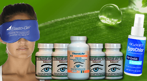
TheraLife, we recommended the following regimen to treat MGD.
- TheraLife Eye revitalizes tear secretion glands, including meibomian glands. –
- Hot compress- twice a day, 10 minutes each time
- Use Thermal Pulse System to melt the clogging and open the secretion glands. The procedure may need to be repeated every 9-12 months.
- Use Intense Light Pulse (IPL) therapy using laser heat to open the glands.
- Use steroid drops to stop inflammation when the condition becomes severe.
Over time, the meibomian glands accumulate dead epithelial cells (just like dead skin cells) around the eyelid, making it impossible for the natural lubricants to flow out.
Follow us on Facebook and Twitter.
Call us toll-free 1-877-917-1989
Frequently Asked Questions
How do you express a clogged meibomian gland?
How do you unclog your eye glands?
- Hot Comoress twice daily to melt the clogging.
- Debridement – scraping along the eyelid margin to get rid of debre.
- IPL- Intense Pulsed Light
- LipiFlow –
- Intraductal Meibomian Probing – for eyes that nothing comes out. This is a surgical procedure.
Please do not attempt to unclog your own oil glands to avoid injury.
How long does it take to clear meibomian glands?
Conclusion
Meibomian gland disease (MGD) is a broad term involving meibomian gland dysfunction that results in altered meibomian gland secretions and tear-film abnormalities. MGD can be congenital or acquired but is more commonly acquired in nature. It is seen more commonly with advancing age but also typical in youngsters, particularly those involved in excessive digital device use, exposed to wind or extreme sunlight conditions, or contact lens users. MGD affects the meibum secretions, which leads to blocked meibomian ducts and thus decreased lipid secretion, altering the tear film composition. The reduced tear film stability results in increased evaporation, hyperosmolarity, inflammation, and ocular surface disorder.
Manually uncloging meibomian glands can facilitate in dry eye recovery and should be considered.
Get TheraLife help when treating dry eyes with eye drops just do not work. Try the clinically tested oral dry eye treatment that works. Contact us now 1-877-917-1989
References
1.Craig JP, Nichols KK, Akpek EK, Caffery B, Dua HS, Joo CK, et al. TFOS DEWS II definition and classification report. Ocul Surf. 2017;15(3):276–83.
2.Alghamdi YA, Mercado C, McClellan AL, Batawi H, Karp CL, Galor A. Epidemiology of meibomian gland dysfunction in an elderly population. Cornea. 2016;35(6):731–5.
3.Lemp MA, Crews LA, Bron AJ, Foulks GN, Sullivan BD. Distribution of aqueous-deficient and evaporative dry eye in a clinic-based patient cohort: a retrospective study. Cornea. 2012;31(5):472–8.
4.Mathers WD. Ocular evaporation in meibomian gland dysfunction and dry eye. Ophthalmology. 1993;100(3):347–51.
5.Nelson JD, Shimazaki J, Benitez-del-Castillo JM, Craig JP, McCulley JP, Den S, et al. The international workshop on meibomian gland dysfunction: report of the definition and classification subcommittee. Invest Ophthalmol Vis Sci. 2011;52(4):1930–7.
6.Schaumberg DA, Uchino M, Christen WG, Semba RD, Buring JE, Li JZ. Patient reported differences in dry eye disease between men and women: impact, management, and patient satisfaction. PLoS One. 2013;8(9):e76121.
7.Shen Lee B, Kabat AG, Bacharach J, Karpecki P, Luchs J. Managing dry eye disease and facilitating realistic patient expectations: a review and appraisal of current therapies. Clin Ophthalmol. 2020;14:119–26.
8.Lee H, Chung B, Kim KS, Seo KY, Choi BJ, Kim TI. Effects of topical loteprednol etabonate on tear cytokines and clinical outcomes in moderate and severe meibomian gland dysfunction: randomized clinical trial. Am J Ophthalmol. 2014;158(6):1172–1183 e1171.
9.Ciloglu E, Ozcan AA, Incekalan T, Unal F. The role of topical azithromycin in the treatment of meibomian gland dysfunction. Cornea. 2020;39(3):321–4.
10.Lee H, Min K, Kim EK, Kim TI. Minocycline controls clinical outcomes and inflammatory cytokines in moderate and severe meibomian gland dysfunction. Am J Ophthalmol. 2012;154(6):949–957 e941.
11.Geerling G, Tauber J, Baudouin C, Goto E, Matsumoto Y, O’Brien T, et al. The international workshop on meibomian gland dysfunction: report of the subcommittee on management and treatment of meibomian gland dysfunction. Invest Ophthalmol Vis Sci. 2011;52(4):2050–64.
12.Xue AL, Wang MTM, Ormonde SE, Craig JP. Randomised double-masked placebo-controlled trial of the cumulative treatment efficacy profile of intense pulsed light therapy for meibomian gland dysfunction. Ocul Surf. 2020.
13.Piyacomn Y, Kasetsuwan N, Reinprayoon U, Satitpitakul V, Tesapirat L. Efficacy and safety of intense pulsed light in patients with meibomian gland dysfunction-a randomized, double-masked, sham-controlled clinical trial. Cornea. 2020;39(3):325–32.
14.Sabeti S, Kheirkhah A, Yin J, Dana R. Management of meibomian gland dysfunction: a review. Surv Ophthalmol. 2020;65(2):205–17.
15.Arita R, Fukuoka S, Morishige N. Therapeutic efficacy of intense pulsed light in patients with refractory meibomian gland dysfunction. Ocul Surf. 2019;17(1):104–10. .
16.Arita R, Mizoguchi T, Fukuoka S, Morishige N. Multicenter study of intense pulsed light therapy for patients with refractory meibomian gland dysfunction. Cornea. 2018;37(12):1566–71.
17.Sambhi RS, Sambhi GDS, Mather R, Malvankar-Mehta MS. Intense pulsed light therapy with meibomian gland expression for dry eye disease. Can J Ophthalmol. 2020;55(3):189–98.
18.Epstein IJ, Rosenberg E, Stuber R, Choi MB, Donnenfeld ED, Perry HD. Double-masked and unmasked prospective study of terpinen-4-ol lid scrubs with microblepharoexfoliation for the treatment of demodex blepharitis. Cornea. 2020;39(4):408–16.
19.Nichols KK, Foulks GN, Bron AJ, Glasgow BJ, Dogru M, Tsubota K, et al. The international workshop on meibomian gland dysfunction: executive summary. Invest Ophthalmol Vis Sci. 2011;52(4):1922–9.
20.Tomlinson A, Bron AJ, Korb DR, Amano S, Paugh JR, Pearce EI, et al. The international workshop on meibomian gland dysfunction: report of the diagnosis subcommittee. Invest Ophthalmol Vis Sci. 2011;52(4):2006–49.
21.Fukuoka S, Arita R. Tear film lipid layer increase after diquafosol instillation in dry eye patients with meibomian gland dysfunction: a randomized clinical study. Sci Rep. 2019;9(1):9091.
22.Rasmussen A, Ice JA, Li H, Grundahl K, Kelly JA, Radfar L, et al. Comparison of the American-European Consensus Group Sjogren’s syndrome classification criteria to newly proposed American College of Rheumatology criteria in a large, carefully characterised sicca cohort. Ann Rheum Dis. 2014;73(1):31–8.
23.Whitcher JP, Shiboski CH, Shiboski SC, Heidenreich AM, Kitagawa K, Zhang S, et al. A simplified quantitative method for assessing keratoconjunctivitis sicca from the Sjogren’s Syndrome International Registry. Am J Ophthalmol. 2010;149(3):405–15.
24.Lee H, Han YE, Park SY, Lee JH, Chung HS, Moon SY, et al. Changes in the expression of matrix metalloproteinase-9 after intense pulsed light therapy combined with meibomian gland expression in moderate and severe meibomian gland dysfunction. Cont Lens Anterior Eye. 2020.
25.Chotikavanich S, de Paiva CS, de Li Q, Chen JJ, Bian F, Farley WJ, et al. Production and activity of matrix metalloproteinase-9 on the ocular surface increase in dysfunctional tear syndrome. Invest Ophthalmol Vis Sci. 2009;50(7):3203–9.
26.Shimazaki J, Sakata M, Tsubota K. Ocular surface changes and discomfort in patients with meibomian gland dysfunction. Arch Ophthalmol. 1995;113(10):1266–70.
27.Korb DR, Blackie CA. Meibomian gland therapeutic expression: quantifying the applied pressure and the limitation of resulting pain. Eye Contact Lens. 2011;37(5):298–301.
28.Rynerson JM, Perry HD. DEBS – a unification theory for dry eye and blepharitis. Clin Ophthalmol. 2016;10:2455–67.
29.Nattis A, Perry HD, Rosenberg ED, Donnenfeld ED. Influence of bacterial burden on meibomian gland dysfunction and ocular surface disease. Clin Ophthalmol. 2019;13:1225–34. .
30. Dong X, Wang Y, Wang W, Lin P, Huang Y. Composition and diversity of bacterial community on the ocular surface of patients with meibomian gland dysfunction. Invest Ophthalmol Vis Sci. 2019;60(14):4774–83.
31.Mireille Aye A, Bonnin-Jusserand M, Brian-Jaisson F, Ortalo-Magne A, Culioli G, Koffi Nevry R, et al. Modulation of violacein production and phenotypes associated with biofilm by exogenous quorum sensing N-acylhomoserine lactones in the marine bacterium Pseudoalteromonas ulvae TC14. Microbiology. 2015;161(10):2039–51.
32.Hastings JW, Greenberg EP. Quorum sensing: the explanation of a curious phenomenon reveals a common characteristic of bacteria. J Bacteriol. 1999;181(9):2667–833.
33.Yarwood JM, Schlievert PM. Quorum sensing in Staphylococcus infections. J Clin Invest. 2003;112(11):1620–5.
34.Salem DA, El-Shazly A, Nabih N, El-Bayoumy Y, Saleh S. Evaluation of the efficacy of oral ivermectin in comparison with ivermectin-metronidazole combined therapy in the treatment of ocular and skin lesions of Demodex folliculorum. Int J Infect Dis. 2013;17(5):e343–7.
35.Gao YY, Di Pascuale MA, Elizondo A, Tseng SC. Clinical treatment of ocular demodecosis by lid scrub with tea tree oil. Cornea. 2007;26(2):136–43.
36.Choi M, Han SJ, Ji YW, Choi YJ, Jun I, Alotaibi MH, et al. Meibum expressibility improvement as a therapeutic target of intense pulsed light treatment in meibomian gland dysfunction and its association with tear inflammatory cytokines. Sci Rep. 2019;9(1):7648. .
37.Afonso AA, Sobrin L, Monroy DC, Selzer M, Lokeshwar B, Pflugfelder SC. Tear fluid gelatinase B activity correlates with IL-1alpha concentration and fluorescein clearance in ocular rosacea. Invest Ophthalmol Vis Sci. 1999;40(11):2506–12.
38.Kaufman HE. The practical detection of MMP-9 diagnoses ocular surface disease and may help prevent its complications. Cornea. 2013;32(2):211–6..
39.Solomon A, Dursun D, Liu Z, Xie Y, Macri A, Pflugfelder SC. Pro- and anti-inflammatory forms of interleukin-1 in the tear fluid and conjunctiva of patients with dry-eye disease. Invest Ophthalmol Vis Sci. 2001;42(10):2283–92.
40.Sambursky R, Davitt WF 3rd, Latkany R, Tauber S, Starr C, Friedberg M, et al. Sensitivity and specificity of a point-of-care matrix metalloproteinase 9 immunoassay for diagnosing inflammation related to dry eye. JAMA Ophthalmol. 2013;131(1):24–8.
41.Sambursky R, Davitt WF 3rd, Friedberg M, Tauber S. Prospective, multicenter, clinical evaluation of point-of-care matrix metalloproteinase-9 test for confirming dry eye disease. Cornea. 2014;33(8):812–8.
42.VanDerMeid KR, Su SP, Ward KW, Zhang JZ. Correlation of tear inflammatory cytokines and matrix metalloproteinases with four dry eye diagnostic tests. Invest Ophthalmol Vis Sci. 2012;53(3):1512–8.
43.Flanagan JL, Khandekar N, Zhu H, Watanabe K, Markoulli M, Flanagan JT, et al. Glycerol monolaurate inhibits lipase production by clinical ocular isolates without affecting bacterial cell viability. Invest Ophthalmol Vis Sci. 2016;57(2):544–50.
44.Souchier M, Joffre C, Gregoire S, Bretillon L, Muselier A, Acar N, et al. Changes in meibomian fatty acids and clinical signs in patients with meibomian gland dysfunction after minocycline treatment. Br J Ophthalmol. 2008;92(6):819–22.
45.Borchman D, Ramakrishnan V, Henry C, Ramasubramanian A. Differences in meibum and tear lipid composition and conformation. Cornea. 2020;39(1):122–8.
46.Knop E, Knop N, Millar T, Obata H, Sullivan DA. The international workshop on meibomian gland dysfunction: report of the subcommittee on anatomy, physiology, and pathophysiology of the meibomian gland. Invest Ophthalmol Vis Sci. 2011;52(4):1938–78.
47.Xiao J, Adil MY, Chen X, Utheim OA, Raeder S, Tonseth KA, et al. Functional and morphological evaluation of meibomian glands in the assessment of meibomian gland dysfunction subtype and severity. Am J Ophthalmol. 2020;209:160–7.
48.Baudouin C, Messmer EM, Aragona P, Geerling G, Akova YA, Benitez-del-Castillo J, et al. Revisiting the vicious circle of dry eye disease: a focus on the pathophysiology of meibomian gland dysfunction. Br J Ophthalmol. 2016;100(3):300–6.