Impact of diets on dry eyes, blepharitis, Meibomian Gland Dysfunction, Ocular Rosacea, and autoimmune Sjogren’s diseases.
Diet and dietary components are known to directly impact the risk factors for dry eyes, blepharitis, meibomian gland dysfunction, and autoimmune Sjogren’s diseases. The chronic diseases that affect the eyes.
The majority of these problems can be averted with timely intervention and the treatment goal is to keep the ocular surface as healthy.
Goal
Our goal is to review what diets work for these dry eye diseases and discuss each disorder in detail.
Recover Faster with TheraLife Dry Eye Treatment and Autoimmune Diet
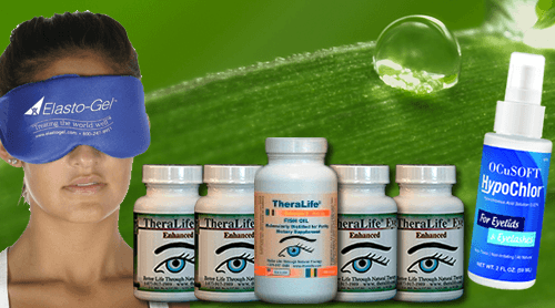
TheraLife deveoped a protocol – combination of Theralife Eye capsules, Omega 3 fish oil, hot compress and Avenova eyelid cleanser that works.
All natural, clinically proven, doctors recommended. Money back guaranteed
- TheraLIfe Eye capsules to restore and revive tear production
- Omega-3 fish oil – anti-inflammatory, lubrication to thicken tears
- Hot Compress – melt the blockages in your ,meibomian oil glands
- Avenova Eyelid Cleanser – eyelid hygiene, get rid of blepharitis
Add To Cart
How Does TheraLife Eye Work?
What is in Theralife Eye Capsules?
Customer Success Stories
Severe Chronic Dry Eyes- ocular rosacea
I have had chronic severe dry eyes for many years I am writing to let you know how thankful I am for your product- TheraLife Eye. It has virtually changed my life. Instead of ALWAYS thinking about my sore, red, dry eyes, I NEVER think about them anymore. And every time I look in the mirror and see my bright, wide, youthful looking eyes I make a little wish that your company is thriving and I will never again have to be without TheraLife Eye capsules. You know, when your eyes are red and you squint in discomfort, you really do look older.
I’ll be traveling to Las Vegas soon and am anxious to see how I do in the desert air. Wish me luck! A bottle of TheraLife Eye will be in my purse for sure.
Thank you, thank you, thank you.
C.L. Ohio, United States
What diets will we review?
We will review a selection of popular diets – including the Mediterranean, Paleo, gluten-free, and a low arachidonic acid diet. We are looking at their effect on digestive health and dry eyes, autoimmune and blepharitis diet.
How do diets work?
Diets demonstrated that including or excluding particular dietary components works to reduce inflammation. A diet based on whole food eating reduces intestinal microbial imbalance, supports intestinal integrity, and modulates inflammation.
What chronic diseases are on the rise?
The number of chronic diseases is growing- including autoimmune disease, cancer, cardiovascular disease, and diabetes, in addition to the ever-growing cost of marginally effective pharmaceuticals. We are reaching a crisis point in chronic disease management. We need to employ rigorous scientific inquiry into cost-effective and readily available dietary and lifestyle therapies.
Why is dietary control of inflammation so difficult?
Dietary control is difficult because of the emphasis on pharmaceuticals and the focus on epidemiologic. Observational studies aimed at assessing dietary patterns in large populations are lacking. Society has suffered tremendously with a lack of well-designed, prevention-directed clinical trials utilizing dietary and lifestyle therapies.
The scientific community is missing data of well-designed, interventionally-directed clinical trials utilizing dietary and lifestyle therapies to address the growing chronic disease management crisis as an alternative to the costly but marginally effective pharmaceuticals. The main factor in this missing data is the lack of funding for conducting large-scale dietary and lifestyle-based interventions. In reviewing recent interventional research examining the role of one such dietary and lifestyle-based intervention, we will discuss the Autoimmune Protocol (AIP) diet below..
Most blepharitis is associated with a staphylococcal bacterial infection on the ocular surface. The irritation is from bacterial toxins and enhanced cell-mediated immunity to Staphylococcus aureus.
Diet has shown to be effective
Dietary modification as an adjunct to IBD therapy.
Increasing evidence suggests that dietary modification can modulate inflammation and improve clinical responses in IBD. A prospective observational study indicates that an AIP diet involving an elimination phase followed by a maintenance phase demonstrates preliminary efficacy in patients with active Irritable Bowel Disease (IBD). 73%of study participants achieved clinical remission, and all maintained clinical remission during the study’s maintenance phase. This dietary study was an adjunct to medical treatment, and almost 50% of patients in the study were on biological therapy. Therefore, dietary modification can be an adjunct to conventional IBD therapy, even with moderate to severe disease.
What are the diet strategies that work?
Dietary strategies, such as:
- limiting arachidonic acid to <90 mg per day, avoiding gluten-containing products,
- consuming fiber, incorporating fermented foods,
- increasing fruit and vegetable intake to above six portions daily
- moderating sodium and long-chain saturated fat consumption.
- OMega 3 fish oil – strongly anti-inflammatory.
Incorporating these dietary aspects may lay the foundations for developing a diet for chronic dry eyes, blepharitis, and autoimmune diseases as a preventive health measure.
Reduce inflammation for Autoimmune Diseases
We focus on the Autoimmune diet to reduce inflammation for chronic inflammation, including blepharitis and autoimmune diseases that affect the eyes. Eye disorders include but are not limited to: chronic blepharitis, ocular rosacea, chronic dry eyes, Iritis, Uveitis. Plus, autoimmune diseases such as Sjogren’s, lupus, rheumatoid arthritis, Hashimoto’s thyroiditis, grave’s disease, graft versus host disease, and more.
What is the Paleo Diet for chronic disorders?
The Paleo diet, which is very similar to Autoimmune Protocol (AIP), is a diet that helps heal the immune system and gut mucosa. It applies to any inflammatory disease. The Autoimmune diet is a subset of the Paleo diet. Sometimes referred to as the “ancestral diet,” the Paleo diet is believed to be like the foods eaten by early humans, emphasizing whole foods, including organic vegetables, fruits, grass-fed and naturally-raised meats, wild-caught fish, eggs, nuts, and seeds. Besides avoiding all processed foods and sugar, it also omits all grains, dairy products, beans, and legumes (peas, including peanuts). Supporters of the Paleo diet recommend eliminating these foods because the human digestive system is not well-equipped to break down these foods. The result is causing digestion issues, allergies, and sensitivities, and inflammation.
Is TheraLife Eye capsule suitable for Paleo Diet/ AIP Diet?
Yes, TheraLife Eye capsules are made entirely of plant-based extracts. Use it at the same time as being on a diet.
The gut is a vital issue with inflammation.
Many of us believe, all autoimmune diseases stem from the gut- low absorption of nutrients. Therefore, probiotics are a must to treat autoimmune diseases and their related inflammation.
Why is it difficult in diagnosing chronic diseases
We have a problem in this country with how we eat, treat disease and heal disease. AIP addresses inflammation in the gut that causes chronic diseases. Autoimmune disease is a condition where the body cannot distinguish between healthy tissue and foreign invaders. A hypersensitive reaction occurs. The body starts attacking its tissue. Attaching one’s tissue can occur silently until a full-blown autoimmune disease develops. All autoimmune diseases have in common is tissue self-attacking. In particular, Sjogren’s attack places like the eyes, mouth, thyroid gland, brain tissue, or salivary glands, to name a few. On average, it takes seven (7) years to diagnose Sjogren’s disease.
How does Paleo Diet Work?
The Paleo diet works to reduce inflammation in the intestines. Paleo works to calm inflammation in the gut and also calm inflammation in the body. And while there is no cure for some chronic diseases like dry eyes, it can go into remission. The Paleo diet targets healing the intestinal mucosa and supporting low inflammation in the body that can be a lifestyle change.
The Autoimmune Diet (AIP)
The Autoimmune Protocol is a version of the Paleo diet that addresses inflammation that causes Autoimmune Disease. This inflammation has its beginning roots in the gut. Diet is one aspect of healing, and it is the most crucial aspect of long-term health.
What diseases does AIP address?
Many divergent paths have a summation with the AIP lifestyle like an autoimmune disease; adrenal fatigue, liver congestion, hormone imbalances, and insulin resistance play a role in using supplements and the AIP diet.
How to make the AIP Diet work?
To be successful, it requires one to be compliant and diligently following the diet. Each person who decides to try the AIP diet should work with a practitioner. This practitioner can help determine if you are candidates for low histamine, low latex, and low FODMAP in addition to following AIP. Other adjunct protocols may include functional blood chemistry, saliva hormone testing, saliva adrenal testing, stool testing, and antibody tests.
What causes Autoimmune Diet not to work?
Before starting the AIP, it is essential to get blood work or other tests like adrenal tests. AIP diet will not work if you have dysglycemia; insulin resistance; anemia (not all anemias are from low iron!); intestinal or other infections like h. pylori; SIBO, h-p axis issues; and adrenal dysfunction.
Why consider an autoimmune diet?
The premise of the AIP diet involves a staged elimination of food groups that may be associated with immune stimulation. Food that can be intolerant, plus maintenance of the eliminated foods. The staged reintroduction of certain foods or food groups over time.
- AIP (an autoimmune disease) is a disease of inflammation that causes attacks on one’s tissue.
- Food is a great way to reduce inflammation and calm the immune system.
- Diet is usually NOT enough. Specific protocols of gut healing and removing SIBO and immune support supplements may be necessary.
- Committed to the diet makes AIP work.
- Avoid food sensitivities in this diet. If you are sensitive to carrots, for example, don’t eat them while on AIP.
- Additional Supplements are usually required to heal the gut fully.
- Allow green beans, snow peas, and sugar snap peas unless low FODMAPS is needed. Because green beans are not mature bean seeds. AIP states no ripe beans seeds from the legume family
- Brain Chemistry and Adrenal Fatigue are likely culprits in autoimmune diseases.
- Undiagnosed Insulin Resistant Hypoglycemia is a significant factor in inflammation that can contribute to autoimmune disease, adrenal fatigue, and brain dis-regulation.
- All of the above-listed foods can be gut irritants, exacerbate dysbiosis in the gut, and contribute to SIBO (small intestine bacterial overgrowth). Eliminating these foods is vital to reducing inflammation. AIP can help address the GALT imbalances (Gut Associated Lymphoid Tissue) in the intestines. The gut contains a large percentage of immune system GALT tissues (70%). The gut mediates the T & B Lymphocytes that carry out immune system attacks by producing antigens or antibodies. The goal of AIP is to reduce this from occurring. Gut-mediated inflammation is applicable for addressing Autoimmune Disease.
AIP Diet PHASE 1: 6-8 Weeks
NOT ALLOWED:
- Nuts (including nut oils like walnut and sesame seed oils)
- Seeds (including flax, chia, pumpkin, sunflower, sesame, and culinary herb seeds like cumin and coriander)
- Beans/Legumes (this includes all beans like kidney, pinto, black as well as Soy in all its forms)
- Grains (Corn, Wheat, Millet, Buckwheat, Rice, Sorghum, Amaranth, Rye, Spelt, Teff, Kamut, Oats, etc.)
- Alternative sweeteners like xylitol and stevia
- Dried fruits and over-consumption of fructose (I recommend up to 2 pieces of fruit a day)
- Dairy Products
- All Processed Foods
- Alcohol
- Chocolate
- Eggs
- Nightshades (tomatoes, potatoes, peppers, eggplant, mustard seeds, all chili’s including spices)
- No vegetable oils (olive oil, lard, cultured ghee, and coconut oils are permitted)
- Culinary herbs from seeds (mustard, cumin, coriander, fennel, cardamom, fenugreek, caraway, nutmeg, dill seed)
- Tapioca. I eliminate this in the first 6-8 weeks because it is a known gluten cross reactor according to Cyrex Labs Gluten Cross-Reactivity Test.
ALLOWED:
- Vegetables (except nightshades)
- Fruits (limit to 15-20 grams fructose/day)
- Coconut products include coconut oil, manna, creamed coconut, coconut aminos, canned coconut milk (with no additives), shredded coconut (this list does not include coconut sugar and nectar)
- Fats: olive oil, coconut oil, avocados, lard, bacon fat, cultured ghee (certified to be free of casein and lactose)
- Fermented Foods (coconut yogurt, kombucha, water and coconut kefir, fermented vegetables)
- Bone Broth
- Grass-Fed Meats, Poultry, and Seafood
- Non-Seed Herbal Teas
- Green Tea
- Vinegar: Apple Cider Vinegar, Coconut vinegar, red wine vinegar, balsamic.
- Sweeteners: occasional and sparse use of honey and maple syrup (1 tsp/day)
- Herbs: all fresh and non-seed herbs are allowed (basil tarragon, thyme, mint, oregano, rosemary, ginger, turmeric, cinnamon, savory, edible flowers)
- Binders: Grass-Fed Gelatin and Arrowroot Starch (watch the starch, however, if you have adrenal issues)
How to reintroduce foods after Phase 1
- 72 Hour Rule: It takes 72 hours to produce an IgA, IgG and IgM mediated antibody symptom. It can be physical or mental. Symptoms such as lethargy, brain fog, aching joints, rashes, stomach aches, numbness, feeling hungover, bloating, gas, constipation, insomnia, fatigue, memory loss are common.
- Reintroduce only one food every five days. When you reintroduce the food, eat enough to elicit a response—a small bite, then a few hours a spoonful, and then that night a serving.
- Keep a food reintroduction notebook. Reintroduce food after a cleanse or AIP, and they have a sensitivity symptom. They can’t remember what or when they did the reintroduction. Writing everything down helps a lot.
Doing yearly or twice yearly cleanses and support phases, eating fermented foods and bone broth at least 2-3 times a week in 1/2 cup portions are very helpful.
What to avoid if you have a chronic disease.
- Eggs (especially the whites)
- Nuts
- Seeds(including cocoa, coffee, and seed-based spices)
- Nightshades(potatoes, tomatoes, eggplants, sweet and hot peppers, cayenne, red pepper, tomatillos, goji berries, etc. and spices derived from peppers, including paprika)
- Potential Gluten Cross-Reactive Foods
- Fructose consumption over 20g per day
- Alcohol
- NSAIDS (like aspirin or ibuprofen)
- Non-nutritive sweeteners (yes, all of them, even stevia)
- Emulsifiers, thickeners, and other food additives
What to eat if you have a chronic disease
- Organ meat and offal (aim for five times per week, the more, the better)–read more.
- Fish and shellfish (wild is best, but farmed is okay) (aim for at least three times per week, the more, the better)–read
- vegetables of all kinds, avoid the nightshades, aim for 8-14 cups per day
- Sea vegetables (excluding algae like chlorella and spirulina, which are immune stimulators)
- quality meats(grass-fed, pasture-raised, wild as much as possible) (poultry in moderation due to high omega-6 content unless you are eating a ton of fish)
- quality fats(pasture-raised/grass-fed animal fats [rendered or as part of your meat], fatty fish, olive, avocado, coconut, palm [not palm kernel])
- fruit (keeping fructose intake between 10g and 20 g daily)
- probiotic foods(fermented vegetables or fruit, kombucha, water kefir, coconut milk kefir, coconut milk yogurt, supplements)
- glycine-rich foods(anything with connective tissue, joints or skin, organ meat, and bone broth
What is FODMAP?
FODMAP stands for fermentable oligosaccharides, disaccharides, monosaccharides, polyols, short-chain carbohydrates (sugars) that the small intestine absorbs poorly. Some people experience digestive distress after eating them. Symptoms include Cramping.
High FODMAPS may disagree with some on the AIP diet. For example, nectarines, coconut, or onions may bother some people.
If you are reacting to certain starches in foods – Avoid high FODMAPS from your diet. If you are FODMAPS sensitive, eliminate for 10-14 days and then slowly reintroduce.
What is Small Intestinal Bacterial Overgrowth (SIBO)
Small intestinal bacterial overgrowth (SIBO) is a severe condition affecting the small intestine. When bacteria that usually grow in other parts of the gut start growing in the small intestine, that causes pain and diarrhea.
Researches in the Role of Diet in Autoimmune Disease:
Gluten in Inflammatory Bowel Disease
Outside of the removal of gluten in celiac disease, a pathology directly linked to an immune reaction to gluten and gluten-self immune complexes, there are inconsistent and often vague dietary and lifestyle recommendations provided to individuals affected by the over 80 recognized autoimmune diseases. To date, dietary and lifestyle-informed research has focused disproportionately on Inflammatory Bowel Disease (IBD). Most other autoimmune disorders lack specific dietary guidelines or rigorous evidence for any particular dietary pattern. Research continues to demonstrate the harmful health effects of a diet high in ultra-processed refined foods. Given the inherent complexity in studying the impact of a single food or nutrient compared to broader nutritional patterns, doctors will likely continue to face challenges in providing rigorous and evidence-based dietary recommendations to both healthy and diseased populations.
Research shows that eliminating intolerant food groups can improve symptoms and endoscopic inflammation in patients with Irritable Bower Disease (IBD). Dietary change can be an essential adjunct to IBD therapy to achieve remission and perhaps enhance the durability of response and remission. Probably for a subset of patients, dietary and lifestyle modification alone may be sufficient to control underlying luminal inflammation. Practitioners should counsel patients wishing to incorporate dietary therapy on options assessed for micronutrient deficiencies and monitored routinely. Integrating and coordinating care with health coaches and registered dieticians can allow for practical education and implementation of dietary modification over time, following unique patient goals.
Leaky Gut Syndrome
A simple prescription of a whole foods diet that minimizes refined foods is likely insufficient for all symptomatic individuals affected with mild to moderately severe autoimmune disease. Those with an autoimmune disorder also consider the nutrient density of specific whole foods. Especially those foods implicated in immune dysregulation. Including foods that disrupt either the gut microbiota ecosystem or the mucosal gut barrier. Therefore, establishing a theoretical basis for the inclusion or exclusion of certain foods.
Autoimmune Protocol (AIP) In IBD and Ulcerative Colitis
This theoretically developed dietary approach, known as the Autoimmune Protocol (AIP), has gained significant anecdotal recognition in a broader clinical community as an effective nutrient-dense diet that can address quality of life and disease activity across a broad spectrum of autoimmune diseases. The research studied the effectiveness of AIP alongside community-based health coaching for 15 individuals with long-standing IBD. The researchers noted that 11/15 participants (5 with Ulcerative Colitis and 6 with Crohn’s Disease) achieved clinical remission by week 6 of the study and maintained remission by study end at week 10. In addition, they noted decreases in the inflammatory marker fecal calprotectin. The study includes changes to the gastrointestinal mucosa as part of follow-up endoscopy or colonoscopy. The subjective and objective changes were rather staggering. They provided the first scientific evidence for the efficacy of the AIP diet for two common autoimmune diseases.
Hashimotos Thyroiditis
The AIP diet for individuals with an autoimmune thyroid disease known as Hashimoto’s Thyroiditis is studied. Seventeen non-obese women aged 20-45 with a history of Hashimoto’s Thyroiditis and no other active autoimmune disease were enrolled. The 17 women participated in a 10-week online, community-based health coaching program implementing the AIP diet. T Following the 10-week program, statistically and clinically significant changes were observed. 6/13 women who began the study on some form of thyroid hormone replacement medication were able to decrease their medication by the end of the study. Statistically significant decreases in self-reported weight and BMI happened. When examining changes in thyroid hormones and thyroid antibodies, there were no clinically or statistically significant changes.
Blepharitis
What is Blepharitis
Blepharitis is the inflammation of the eyelids. Symptoms of blepharitis include crusty, red, itchy eyelids.
Blepharitis, an inflammatory condition of the eyelid margin, is a common cause of ocular discomfort and irritation in all age and ethnic groups. Blepharitis can lead to permanent changes in the eyelid margin or vision loss -including superficial keratopathy, corneal neovascularization, and ulceration.
What causes blepharitis?
The root cause of blepharitis is chronic dry eye. There are two types of blepharitis:
- Anterior blepharitis – bacterial infection or mites
- Posterior blepharitis- results in clogging of the meibomian oil glands (MGD). More discussions later.
Blepharitis Symptoms
Your eyes may appear red, and your eyelids may appear oily, swollen, and red, flaking the skin around your eyelashes with crust formation.
What is the difference between anterior and posterior blepharitis?
Anterior blepharitis refers to inflammation mainly centered around the skin, eyelashes, and lash follicles. At the same time, the posterior variant involves the meibomian oil gland orifices, meibomian glands, tarsal plate, and blepharo-conjunctival junction.
Can you have both anterior and posterior blepharitis?
Blepharitis may be asymptomatic. However, when present, the symptoms of anterior blepharitis, posterior blepharitis, and mixed anterior and posterior blepharitis are similar: ocular discomfort, soreness, burning, itching.
What makes blepharitis chronic?
Posterior blepharitis, chronic dry eyes result in MGD. All three conditions set up chronic inflammation, which can become a vicious cycle.
Ocular rosacea, a chronic condition, makes chronic blepharitis and dry eyes even harder to treat—more discussion below Consult TheraLife for effective and natural solutions.
There are two types of blepharitis.
- anterior blepharitis
- posterior blepharitis
Anterior blepharitis and posterior blepharitis differ on their anatomic location. However, there is considerable overlap, and both are often present.
Anterior Blepharitis
Anterior blepharitis affects the eyelid skin, base of the eyelashes, and eyelash follicles. It includes the traditional classifications of staphylococcal and seborrheic blepharitis.
Posterior Blepharitis
Posterior blepharitis affects the meibomian oil glands and gland orifices. It has a range of potential etiologies, the primary cause being MGD.
What causes blepharitis?
Staphylococcus bacterial infection
We all have some bacteria on our skin called “normal flora. An overgrowth of the Staphylococcus bacteria on your eyelids and at the base of your eyelashes can result in blepharitis.
What are the other causes of blepharitis?
Other conditions that can contribute to the development of blepharitis include:
- dandruff,
- Mites – infection of eyelids with mites called Demodex.
- Styes and Chalazion – Blepharitis can become chronic and result in recurrent infections of the oil glands. These infections are styes (hordeolum), chalazia (plural of chalazion), and cellulitis.
What helps to control blepharitis?
Bacterial infection and inflammation contribute to blepharitis pathology. Long-term management of symptoms may include daily eyelid cleansing routines and the use of therapeutic agents that reduce infection and inflammation.
How does TheraLife help in treating chronic blepharitis?
The most successful approach is TheraLife. TheraLife treats chronic blepharitis by treating dry eyes, MGD all at the same time. To learn more.
Epidemiology of Blepharitis
Blepharitis is one of the most common ocular disorders encountered in eye doctor’s offices. 37% to 47% of patients seen in the eye doctor’s office have signs of blepharitis.
Blepharitis Risk Factors & Associated Conditions
Dry Eye
Dry eye is present in 50% of patients with staphylococcal blepharitis. In a study of 66 patients with dry eye- 75% had staphylococcal conjunctivitis or blepharitis. Since a decrease in local lysozyme and immunoglobulin levels associated with tear deficiency might alter resistance to bacteria. Therefore, predisposing people to the development of staphylococcal blepharitis.
Approximately 25% to 40% of patients with seborrheic blepharitis and MGD also have dry eye. Dry eyes cause increased tear film evaporation due to a deficiency in the lipid component of the tears as well as reduced ocular surface sensation.
Dermatologic Conditions
Facial Rosacea
20% to 42% of people with all types of blepharitis have acne rosacea on the face. The skin on the nose and cheeks has erythema, telangiectasias, papules, pustules, and prominent sebaceous glands. Severe cases of both acne rosacea and blepharitis can lead to severe eyelash and eyelid damage.
Seborrheic dermatitis
Seborrheic dermatitis has flaking and greasy skin on the scalp, retro auricular area, glabella, and nasolabial folds. Research indicates 33% to 46% of patients with blepharitis have seborrheic dermatitis. In one study, 95% of patients with seborrheic blepharitis also had seborrheic dermatitis.
Demodex- Mites
Demodex infestation, characterized by cylindrical dandruff or sleeves around the eyelashes, has been found in 30% of patients with chronic blepharitis. The infestation and waste of the mites cause blockage of the follicles and glands and inflammatory response. Nonetheless, patients with recalcitrant blepharitis have responded to therapy directed at eradicating the mites with tea tree oil.
Pathophysiology of Blepharitis
The exact pathogenesis of blepharitis is suspected to be multifactorial.
Bacterial infection.
Most blepharitis is associated with a staphylococcal bacterial infection on the ocular surface. The irritation is from bacterial toxins and enhanced cell-mediated immunity to Staphylococcus aureus. In one study of ocular flora, 46% to 51% of those diagnosed with staphylococcal blepharitis had cultures positive for Staphylococcus aureus compared to 8% of normal patients.
Seborrheic dermatitis
Seborrheic blepharitis has less inflammation than staphylococcal blepharitis. Seborrheic blepharitis has more oily or greasy scaling. Some patients with seborrheic blepharitis also exhibit characteristics of MGD.
MGD is the abnormalities of the meibomian glands and altered secretion of the meibum. The meibum plays an essential role in slowing the evaporation of tear film and smoothing the tear film to provide an even optical surface. Both quantitative deficiencies in meibum or qualitative differences in its composition can contribute to symptoms experienced in MGD blepharitis.
How Is Blepharitis Diagnosed?
The diagnosis of blepharitis requires a thorough medical history, listening to your symptoms, and examining your eyes with a slit lamp microscope- for eyelids, eyelid margins, eyelashes, and meibomian gland orifices (openings). Gentle digital pressure is applied to your eyelids to assess the quality and quantity of the oils secreted by your meibomian gland.
The gold standard to directly evaluate your meibomian oil glands structurally is to image them using advanced technology known as LipiScan. LipiScan is a meibographer that captures images of your meibomian glands.
Blepharitis, MGD, and Ocular Rosacea are closely linked.
Blepharitis, meibomian gland dysfunction, and ocular rosacea are closely related conditions with the same treatment protocols.
Blepharitis Physical Examination
While the clinical features of the blepharitis categories can overlap, sure signs and symptoms are more commonly associated with particular subtypes.
Staphylococcal blepharitis
Staphylococcal blepharitis has erythema and edema of the eyelid margin. People may exhibit eyelash loss and misdirection – signs that do now exist in other types of blepharitis. Other symptoms may include telangiectasia on the anterior eyelid, hard scales/collarettes encircling the lash base, and corneal changes (infiltrates, phlyctenules). In severe and long-standing cases, eyelid ulceration and corneal scarring may occur.
Seborrheic blepharitis
Seborrheic blepharitis has less erythema, edema, and telangiectasia of the lid margins than staphylococcal blepharitis. However, Sehorrheic blepharitis has an increased amount of oily scale and greasy crusting on the lashes.
Posterior blepharitis
Posterior blepharitis/MGD, often associated with ocular rosacea, is seen clinically by examining the posterior eyelid margin. The meibomian glands may appear capped with oil, be dilated, or obstructed. The secretions of the glands are usually turbid and thicker than usual. Telangiectasias and lid scarring may also be present in this area. Chalazia may be a cause or consequence of MGD.
In all forms of blepharitis, examination of the tear film may show instability and rapid evaporation. The method most frequently used to assess tear film stability measures the tear breakup time (TBUT), i.e., the time interval between a complete blink and the first appearance of a dry spot in the pre-corneal tear film after fluorescein instillation. There is general agreement that a TBUT shorter than 10 seconds reflects tear film instability.
Blepharitis Treatment – Eyelid Hygiene:
- Warm compress twice each day, in the morning and the evening. You should apply a warm compress to your closed eyes for 10-20 minutes to loosen/soften the oils in the glands of your eyelids (meibomian oil glands). Gel packs or a heat mask such as an Optase or Bruder are heated in a microwave. Electric Heated Dry Eye Mask also works. Washcloths do not work well.
- Massage your eyelids after a warm compress to bring up the clogging.
- Eyelid Cleaning – This should be followed immediately by cleaning your eyelids. We highly recommend Avenova.
We highly recommend the comprehensive Theralife protocol for treating blepharitis, including eyelid hygiene plus dry eyes and MGD. To learn more
What is Meibomian gland dysfunction?
Meibomian gland dysfunction (MGD) is a widespread eye problem. It is inflammation and clogging of tiny glands in your eyelids that produce the oily layer of your tears. When these meibomian glands get inflamed and clog up, less of this oil (meibum) is secreted into the tear film – causing your tears to evaporate more quickly, resulting in dry, irritated eyes.
Symptoms of MGD
MGD is characterized by functional abnormalities of the meibomian oil glands and altered secretion of meibum, which plays a vital role in slowing the evaporation of tear film and smoothing the tear film to provide an even optical surface. Both quantitative deficiencies in meibum or qualitative differences in its composition can contribute to symptoms experienced in this meibomian gland disease.
In addition to dry eyes, symptoms of meibomian gland dysfunction (MGD) include:
- Red eyes
- Itchy eyes
- Burning eyes
- Watery eyes
- Sensitivity to light
- Feeling something is “in” your eye
- Intermittent blurred vision
MGD commonly occurs with an eyelid problem called blepharitis, which causes inflamed eyelids and a crusty discharge at the base of the eyelashes.
Styes also frequently occur with MGD, causing a sensitive red bump at the eyelid margin. And sometimes, MGD can cause a painless bump inside the eyelid – called a meibomian cyst or chalazion.
MGD risk factors
Aging
Aging increases your risk of dry eyes and MGD. A study of 233 older adults (average age 63) found that 59% had at least one sign of MGD.
Ethnic background
Your ethnic background also plays a role. Research has shown up to 69% of Asian populations in Thailand, Japan, and China have meibomian gland dysfunction. By comparison, only up to 20% of non-Hispanic whites in the US and Australia have this meibomian gland disease.
Eye makeup
Wearing eye makeup is another contributing cause of MGD. Eyeliner and other cosmetics can clog the openings of meibomian glands – especially if you don’t thoroughly clean your eyelids and remove all traces of eye makeup before sleep.
Contact Lenses
Wearing contacts may aggravate meibomian gland dysfunction. Research has shown that contact lens wear is associated with meibomian gland changes. These changes remain for up to six months after the use of contact lenses is discontinued.
How is MGD detected?
The symptoms of meibomian gland dysfunction are nearly the same as those of dry eye syndrome. Only an eye doctor can tell for sure if you have MGD.
A simple technique your doctor might use to detect MGD is to apply pressure to your eyelid – to express the contents of the meibomian glands. Observing these secretions often can enable trained eye care professional to determine if you have meibomian gland dysfunction.
Because meibomian gland dysfunction affects the stability of the tear film, your eye doctor also may test the quality, quantity, and stability of your tears.
Tear Breakup Test (TBUT)
One common test is called the tear breakup time (TBUT) test. This simple, painless procedure involves applying a small amount of dye to the tear film on the front surface of your eye. Your doctor then examines your eye with a cobalt blue light, which causes your dyed tears to glow – enabling your eye doctor to see how quickly your tear film loses its stability (breaks up) on the surface of your eye.
Treatments for meibomian gland dysfunction
A common treatment for MGD is applying warm compresses to the eyelids, followed by massaging the eyelids. The goal of this treatment is to unclog the openings of the meibomian glands.
Eye doctors recommend using a specially designed eye mask to deliver heat to the eyelids. The heat therapy to the eyelids is followed by massaging the eyelids to expel the melted oils from the glands.
Unfortunately, warm compresses and lid massages are often insufficient to treat meibomian gland dysfunction and eliminate symptoms.
Other treatments for MGD besides warm compress
When warm compress fails, there are other more advanced treatments for MGD. LipidFlow, IPL, and intraductal meibomian probing, for example.
Autoimmune Diseases
Sjogren’s syndrome
Sjogren’s syndrome is an autoimmune disease. Your immune system attacks parts of your own body by mistake. In Sjogren’s syndrome, it attacks the glands that make tears and saliva – causing a dry mouth and dry eyes. You may have dryness in other places that need moisture, such as your nose, throat, and skin. Sjogren’s can also affect other body parts, including your joints, lungs, kidneys, blood vessels, digestive organs, and nerves.
Most people with Sjogren’s syndrome are women. It usually starts after age 40 -sometimes linked to other diseases such as rheumatoid arthritis and lupus.
Ocular Rosacea
Ocular eye rosacea is inflammation that causes redness, burning, and itching of the eyes. It often develops in people who have acne rosacea, a chronic skin condition that affects the face. Sometimes ocular (eye) rosacea is the first sign that you may later develop the facial type.
Ocular eye rosacea primarily affects adults between the ages of 30 and 50. It seems to develop in people who tend to blush and flush easily.
There’s no cure for ocular rosacea, but medications and a good eye care routine can help control the signs and symptoms.
Symptoms
Signs and symptoms of ocular rosacea can precede the skin symptoms of rosacea, develop at the same time, develop later or occur on their own. Signs and symptoms of ocular rosacea may include:
- Red, burning, itchy or watering eyes
- Dry eyes
- Grittiness or the feeling of having a foreign body in the eye or eyes
- Blurred vision
- Sensitivity to light (photophobia)
- Dilated small blood vessels on the white part of the eye that are visible when you look in a mirror
- Red, swollen eyelids
- Recurrent eye or eyelid infections, such as pink eye (conjunctivitis), blepharitis, sties, or chalazia
The severity of ocular rosacea symptoms doesn’t always match the severity of skin symptoms.
Risk factors
Ocular rosacea is common in people with skin rosacea. However, you can also have ocular rosacea without skin involvement. Skin rosacea affects more women than men, and ocular rosacea affects men and women equally. It’s also more common in fair-skinned people of Celtic and Northern European origin.
Complications
Ocular rosacea may affect the surface of your eye (cornea), particularly when you have dry eyes from the evaporation of tears. Corneal complications can lead to visual symptoms. Inflammation of your eyelids (blepharitis) can cause secondary irritation of the cornea from misdirected eyelashes or other complications. Ultimately, corneal complications can lead to vision loss.
Frequently Asked Questions
What autoimmune disease causes very dry eyes?
Diseases such as Sjogren’s, Rheumatoid Arthritis, Lupus can cause very dry eyes. Seek help from your dry eye specialists. TheraLIfe can also help you alieviate dry eye symptoms together with diet.
What foods trigger dry eyes?
Food that trigger dry eyes include spicy food, food that cause inflamation like brightly colored vegetables and fruits, food with high level of salicylates. Foods with high levels of sugar also can cause dry eyes.
Can an anti inflammatory diet help dry eyes?
References
- Raghupathi W, Raghupathi V. An Empirical Study of Chronic Diseases in the United States: A Visual Analytics Approach. Int J Environ Res Public Health. 2018;15(3):431.
- Knight-Sepulveda K, Kais S, Santaolalla R, Abreu MT. Diet and Inflammatory Bowel Disease. Gastroenterol Hepatol (NY). 2015. 11(8):511– 520.
- Costa CS, Del-Ponte B, Assunção MCF, Santos IS. Consumption of ultra- processed foods and body fat during childhood and adolescence: a systematic review. Public Health Nutr . 2018;21(1):148 159.
- Tapsell LC, Neale EP, Satija A, Hu FB. Foods, Nutrients, and Dietary Patterns: Interconnections and Implications for Dietary Guidelines.Adv Nutr 2016;7(3):445 -454.
- Ballantyne S. The Paleo Approach: Reverse Autoimmune Disease and Heal Your Body. Victory Belt Publishing. Las Vegas, US. 2014.
- Konijeti GG, Kim N, Lewis JD, et al. Efficacy of the autoimmune protocol diet for inflammatory bowel disease. Inflamm Bowel Dis. 2017. 23(11):2054– 2060.
- Abbott RD, Sadowski A, Alt AG. Efficacy of the Autoimmune Protocol Dietas Part of a Multi-disciplinary, Supported Lifestyle Intervention forHashimoto’s Thyroiditis. Cureus. 2019. 11(4).
- Jostins L, Ripke S, Weersma RK, et al. Host-microbe interactions have shaped the genetic architecture of inflammatory bowel disease. Nature. 2012;491:119–124.
- Liu JZ, van Sommeren S, Huang H, et al. Association analyses identify 38 susceptibility loci for inflammatory bowel disease and highlight shared genetic risk across populations. Nat Genet.2015;47:979–986.
- Vindigni SM, Zisman TL, Suskind DL, et al. The intestinal microbiome, barrier function, and immune system in inflammatory bowel disease: a tripartite pathophysiological circuit with implications for new therapeutic directions. Therap Adv Gastroenterol.2016;9:606–625.
- Lewis JD, Abreu MT. Diet as a trigger or therapy for inflammatory bowel diseases. Gastroenterology. 2017;152:398–414.e6.
- Chiba M, Abe T, Tsuda H, et al. Lifestyle-related disease in Crohn’s disease: relapse prevention by a semi-vegetarian diet. World J Gastroenterol.2010;16:2484–2495.
- Obih C, Wahbeh G, Lee D, et al. Specific carbohydrate diet for pediatric inflammatory bowel disease in clinical practice within an academic IBD center. Nutrition. 2016;32:418–425.
- Suskind DL, Wahbeh G, Cohen SA, et al. Patients perceive clinical benefit with the specific carbohydrate diet for inflammatory bowel disease. Dig Dis Sci.2016;61:3255–3260.
- Suskind DL, Cohen SA, Brittnacher MJ, et al. Clinical and fecal microbial changes with diet therapy in active inflammatory bowel disease. J Clin Gastroenterol.[Published online ahead of print December 27, 2016.
- Olendzki BC, Silverstein TD, Persuitte GM, et al. An anti-inflammatory diet as treatment for inflammatory bowel disease: a case series report. Nutr J.2014;13:5.
- Ballantyne S. The Paleo Approach: Reverse Autoimmune Disease and Heal Your Body. Las Vegas: Victory Belt Publishing; 2014.
- Trescott M, Alt A. The Autoimmune Wellness Handbook: A DIY Guide to Living Well with Chronic Illness. New York: Rodale Books; 2016.
- Chassaing B, Van de Wiele T, De Bodt J, et al. Dietary emulsifiers directly alter human microbiota composition and gene expression ex vivo potentiating intestinal inflammation. Gut. 2017;66:1414–1427.
- Martinez-Medina M, Denizot J, Dreux N, et al. Western diet induces dysbiosis with increased E coli in CEABAC10 mice, alters host barrier function favouring AIEC colonisation. Gut. 2014;63:116–124.
- Nickerson KP, Chanin R, McDonald C. Deregulation of intestinal anti-microbial defense by the dietary additive, maltodextrin. Gut Microbes.2015;6:78–83.
- Ghosh S, Louis E, Beaugerie L, et al. Development of the IBD disk: a visual self-administered tool for assessing disability in inflammatory bowel diseases. Inflamm Bowel Dis.2017;23:333–340.
- SAD to AIP in 6. The Gentle Approach to the Autoimmune Protocol. 2016. Available at: at http://www.sadtoaip.com. Accessed December 19, 2015.
- Levesque BG, Sandborn WJ, Ruel J, et al. Converging goals of treatment of inflammatory bowel disease from clinical trials and practice. Gastroenterology. 2015;148:37–51.e1.
- Bernstein CN, Singh S, Graff LA, et al. A prospective population-based study of triggers of symptomatic flares in IBD. Am J Gastroenterol.2010;105:1994–2002.
- Ananthakrishnan AN, Khalili H, Konijeti GG, et al. Sleep duration affects risk for ulcerative colitis: a prospective cohort study. Clin Gastroenterol Hepatol.2014;12:1879–1886.
- Khalili H, Ananthakrishnan AN, Konijeti GG, et al. Physical activity and risk of inflammatory bowel disease: prospective study from the Nurses’ Health Study cohorts. BMJ. 2013;347–f6633.
- Zittan E, Kelly OB, Kirsch R, et al. Low fecal calprotectin correlates with histological remission and mucosal healing in ulcerative colitis and colonic Crohn’s disease. Inflamm Bowel Dis.2016;22:623–630.
30.Huggins AB, Carrasco JR, Eagle RC. MEN 2B masquerading as chronic blepharitis and euryblepharon. Orbit. 2019 Dec;38(6):514-518.
31.Rodriguez-Garcia A, Loya-Garcia D, Hernandez-Quintela E, Navas A. Risk factors for ocular surface damage in Mexican patients with dry eye disease: a population-based study. Clin Ophthalmol. 2019;13:53-62.
32.Choi FD, Juhasz MLW, Atanaskova Mesinkovska N. Topical ketoconazole: a systematic review of current dermatological applications and future developments. J Dermatolog Treat. 2019 Dec;30(8):760-771.
33.Ozkan J, Willcox MD. The Ocular Microbiome: Molecular Characterisation of a Unique and Low Microbial Environment. Curr Eye Res. 2019 Jul;44(7):685-694.
34.Khoo P, Ooi KG, Watson S. Effectiveness of pharmaceutical interventions for meibomian gland dysfunction: An evidence-based review of clinical trials. Clin Exp Ophthalmol. 2019 Jul;47(5):658-668.
35.Soh Qin R, Tong Hak Tien L. Healthcare delivery in meibomian gland dysfunction and blepharitis. Ocul Surf. 2019 Apr;17(2):176-178.
36.Fromstein SR, Harthan JS, Patel J, Opitz DL. Demodex blepharitis: clinical perspectives. Clin Optom (Auckl). 2018;10:57-63.
37.Pflugfelder SC, Karpecki PM, Perez VL. Treatment of blepharitis: recent clinical trials. Ocul Surf. 2014 Oct;12(4):273-84.
38.Kanda Y, Kayama T, Okamoto S, Hashimoto M, Ishida C, Yanai T, Fukumoto M, Kunihiro E. Post-marketing surveillance of levofloxacin 0.5% ophthalmic solution for external ocular infections. Drugs R D. 2012 Dec 01;12(4):177-85.
39.Veldman P, Colby K. Current evidence for topical azithromycin 1% ophthalmic solution in the treatment of blepharitis and blepharitis-associated ocular dryness. Int Ophthalmol Clin. 2011 Fall;51(4):43-52.
40.Hosseini K, Bourque LB, Hays RD. Development and evaluation of a measure of patient-reported symptoms of Blepharitis. Health Qual Life Outcomes. 2018 Jan 11;16(1):11.


