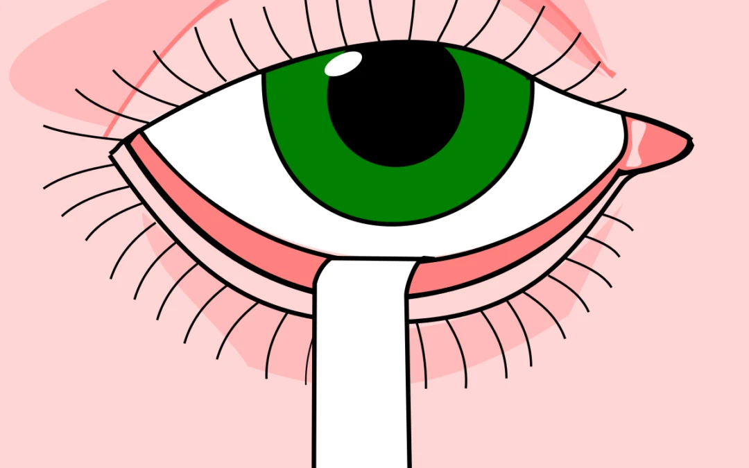
The Schirmer test is to measure tear volume production. Lacrimal glands produce tear-located at the upper outer corner of your eyes. For your eyes to feel comfortable, you need sufficient tear plus tear quality.
How Schirmer’s Test Works for Diagnosis of Dry Eyes
To measure tear volume, the primary test for dry eyes is Schirmer’s Test. Schirmer’s test is performed by putting a topical anesthetic and then placing a thin filter paper in the inferior cul-de-sac. Soft tissue paper can wick up all liquid by capillary attraction before placing the paper strip. The patients’ eyes are then closed for 5 minutes, and the amount of wetting in the paper strip is measured. Less than 5 mm of wetting is abnormal; 5-10 mm is normal.
.The Schirmer I test, without anesthetics, measures both basic and reflex tearing; less than 10 mm of wetting after 5 minutes is diagnostic of dry eyes.
The Schirmer II test, which measures reflex tearing, may be done if the initial Schirmer test yields abnormal results. It is essentially similar to the basic secretion test but with the addition of nasal mucosal irritation induced with a cotton tip applicator. Wetting of less than 15 mm after 5 minutes is consistent with abnormalities of reflex secretion.
The absence of nasal lacrimal reflex tearing, presence of serum autoantibodies, and severe ocular surface disease demonstrated by rose bengal or fluorescein staining argue strongly in favor of a diagnosis of Sjogren’s Syndrome-associated dry eye disease.
Most eye doctors prefer modifying the Schirmer I test to measure basal tear secretion with an anesthetic.
Schirmer’s Test With or Without Anesthetics for Dry Eyes
The Schirmer test has its place in dry eyes clinical care. One of its strengths, aside from being the key discriminating factor between the aqueous-deficient and evaporative dry eyes. It can gain insight into some knowledge about the function of a patient’s lacrimal glands.
To anesthetize or not to anesthetize? Some argue that an anesthetic is required to prevent reflex tearing; others contend that the capacity to measure reflex tears.
The right approach, though, may vary depending on the person. If you want to know if the lacrimal glands can produce aqueous tears as a means to differentiate among dry eye subtypes, then Schirmer without anesthesia would be preferred. If you want to reduce reflex tearing and measure basal tear production, Schirmer with anesthesia may be preferable. Basal tear production is stimulated by the activation of cold fibers on the ocular surface as tears evaporate from the eye. Basal tear production is affected when one has suppressed sensory fibers.
Schirmer’s Test – Open or Closed Eyes?
Should Schirmer’s test be done with open or closed eyes? How to do Schirmer’s test depends on the differences in interpretation. Suppose the eyes are open (with regular blinking) for the patient to casually look at a large 20/400 target. In that case, there will be more reflex tearing, which may be your intent. The ocular surface will also be exposed and could theoretically stimulate some desired basal tear production.
If the eyes are closed, however, basal and reflex production would be decreased. The test then becomes more of a measure of tear volume on the ocular surface than a measure of tear production.
Most eye doctors opt to perform Schirmer without anesthesia (to assess tear production capacity). Doing Schirmer’s test with a closed eyelid is preferred.
Dry Eye Treatment- TheraLife can help
TheraLife Eye capsules is an oral dry eye treatment for long lasting dry eyes relieve all day long
It is all natural, patented, clinical trial proven oral dry eyes treatment that works.
Get started with the discounted Chronic Dry Eye Starter Kit.
Find out how it works – click here
Find out what is in the TheraLife Eye capsules.
References
- Comparison of the clinical effects of carbomer-based lipid-containing gel and hydroxypropyl-guar gel artificial tear formulations in patients with dry eye syndrome: a 4-week, prospective, open-label, randomized, parallel-group, noninferiority study.
Wang TJ, Wang IJ, Ho JD, Chou HC, Lin SY, Huang MC.Clin Ther. 2010 Jan;32(1):44-52. doi: 10.1016/j.clinthera.2010.01.024.PMID: 20171410 Clinical Trial.
- Tear meniscus changes during cotton thread and Schirmer testing.
Yokoi N, Kinoshita S, Bron AJ, Tiffany JM, Sugita J, Inatomi T.Invest Ophthalmol Vis Sci. 2000 Nov;41(12):3748-53.PMID: 11053272
Isreb MA, Greiner JV, Korb DR, Glonek T, Mody SS, Finnemore VM, Reddy CV.Eye (Lond). 2003 Jan;17(1):79-83. doi: 10.1038/sj.eye.6700224.PMID: 12579175
- [The dry eye. Status determination and prospects].
Göbbels M.Fortschr Ophthalmol. 1990;87 Suppl:S190-7.PMID: 2083902 Review. German.
- [Dry eye syndrom–multispecialistic disease. Part two: diagnostic procedure and treatment].
Nowak M, Marek B, Kajdaniuk D, Siemińska L, Kos-Kudła B, Nowak K, Głogowska-Szelag J.Wiad Lek. 2011;64(1):49-55.PMID: 21812364 Review. Polish.
- Reliability and efficacy of maximum fluorescein tear break-up time in diagnosing dry eye disease.
Mou Y, Xiang H, Lin L, Yuan K, Wang X, Wu Y, Min J, Jin X.Sci Rep. 2021 Jun 1;11(1):11517. doi: 10.1038/s41598-021-91110-9.PMID: 34075199 Free PMC article.
Narnoli P, Dhasmana R, Khanduri R.Indian J Ophthalmol. 2021 May;69(5):1178-1182. doi: 10.4103/ijo.IJO_976_20.PMID: 33913855 Free PMC article.
Goh Y, Kwan Z, Han WH, Iqbal T, Yahya F, Khang TF, Singh S.Int Ophthalmol. 2021 Jun;41(6):2139-2147. doi: 10.1007/s10792-021-01771-8. Epub 2021 Mar 31.PMID: 33788072
Kang MS, Shin J, Kwon JM, Huh J, Lee JE.PLoS One. 2021 Jan 11;16(1):e0245329. doi: 10.1371/journal.pone.0245329. eCollection 2021.PMID: 33428686 Free PMC article. Clinical Trial.
Binotti WW, Bayraktutar B, Ozmen MC, Cox SM, Hamrah P.Eye Contact Lens. 2020 Mar;46 Suppl 2(Suppl 2):S84-S105. doi: 10.1097/ICL.0000000000000684.PMID: 31833999 Free PMC article. Review.
- Report of the international dry eye workshop (DEWS). Ocul Surf. 2007;5–2:65–204.
- Schiffman RM, Christianson MD, Jacobsen G, et al. Reliability and validity of the ocular surface disease index. Arch Ophthalmol. 2000;118:615–621.
- Begley CB, Caffrey B, Chalmers RL, et al. Use of the dry eye questionnaire to measure symptoms of ocular irritation in patients with aqueous deficient dry eye. Cornea. 2002;21:664–670
- Rajapopalan K, Abetz L, Mertzanis P, et al. Comparing the discrimative validity of two generic and one disease-specific health-related quality of life measures in a sample of patients with dry eye. Value Health. 2005;8:68–74.
- Korb DR. Survey of preferred tests for diagnosis if tear film and dry eye. Cornea. 2000;19:483–486. doi: 10.1097/00003226-200007000-00016.
- Clinch TE, Benedetto DA, Felberg NT, et al. Schirmer’s test. A closer look. Arch Ophthalmol. 1983;101:1383–1386. [
- Afonso AA, Monroy D, Stern ME, et al. Correlation of tear clearance and Schirmer test scores with ocular irritation symptoms. Ophthalmol. 1999;106:803–810. [
- Loran DFC, French CN, Lam SY, et al. Reliability of the wetting value of tears. Ophthal Physiol Opt. 1987;7:53–56.
- Jordan A, Baum J. Basic tear flow – does it exist? Ophthalmol. 1980;87:920–930
- Josephson JE, Caffrey BE. Corneal staining after instillation of topical anesthetic (SSII) Invest Ophthalmol Vis Sci. 1988;29:1096–1099.
- Cho P. Schirmer Test. I. A Review. Optom Vis Sci. 1993;70(2):152–156. doi: 10.1097/00006324-199302000-00011. [
- Cho P. Schirmer Test. II. A Clinical Study of its Repeatability. Optom Vis Sci. 1993;70(2):157–159. doi: 10.1097/00006324-199302000-00012.
- Nichols KK, Mitchell GL, Zadnik K. The repeatability of clinical measurements of dry Eye. Cornea. 2004;23:272–285. doi: 10.1097/00003226-200404000-00010.
- Kallarackal GU, Ansari EA, Amos N, et al. A comparative study to assess the clinical use of fluorescein meniscus time (FMT) with tear break up time (TBUT) and Schirmer’s tests (ST) in the diagnosis of dry eye. Eye. 2002;16:594–600. doi: 10.1038/sj.eye.6700177.
- Saleh TA, Bates AK, Ewings P. Phenol red thread test vs Schirmer’s test: a comparative study. Eye. 2006;20:913–915. doi: 10.1038/sj.eye.6702052.
- Tomlinson A, Blades KJ, Pearce EI. What does the phenol red thread test actually measure? Optom Vis Sci. 2001;78:142–146. doi: 10.1097/00006324-200103000-00005.
- Nelson PS. A shorter Schirmer test. Optom Mon. 1982;73:568–569.
- Bawazeer AM, Hodge WG. One minute Schirmer test with anesthetic. Cornea. 2003;22:285–287. doi: 10.1097/00003226-200305000-00001. [
- Methodologies to diagnose and monitor dry eye disease: Report of the diagnostic methodology subcommittee on the international dry eye workshop. Ocul Surf 2007;5:108–23.
- Sakamoto R, et al. The phenol red thread tear test: A cross cultural study. Invest Ophthalmol Vis Sci. 1993;34:3510–3514.
- Saleh TA, et al. Phenol red thread test vs. Schirmer’s test: A comparative study. Eye. 2006;20:913–915. doi: 10.1038/sj.eye.6702052.
- Cho P. The cotton thread test: A brief review and a clinical study of its reliability on Hong Kong-Chinese. Optom Vis Sci. 1993;70:804–808. doi: 10.1097/00006324-199310000-00004. [PubMed]
- Patel S, Farrell J, Blades KJ, et al. The value of a phenol red impregnated thread for differentiating between the aqueous and non-aqueous deficient dry eye. Ophthal Physiol Opt. 1998;8:471–476. doi: 10.1016/S0275-5408(98)00005-2.
- Blades KJ, Patel S. The dynamics of tear flow within a phenol red impregnated thread. Ophthal Physiol Opt. 1996;16:409–415. doi: 10.1016/0275-5408(96)00006-3.
- Chiang B, Asbell PA, Franklin B. Phenol-red thread test and Schirmer test for tear production in normal and dry eye patients. Invest Ophthalmol Vis Sci. 1988;29S:337.
- Nichols KK, Nichols JJ, Mitchell GL. The relation between tear film tests in patients with dry eye disease. Ophthal Physiol Opt. 2003;23:553–560. doi: 10.1046/j.1475-1313.2003.00153.x
- Doughty MJ, Whyte J, Li W. The phenol red thread test for lacrimal volume – does it matter if the eyes are open or closed? Ophthal Physiol Opt. 2007;27:482–489. doi: 10.1111/j.1475-1313.2007.00500.x.
- Hurwitz JJ, Maisey MN, Welham RA. Quantitative lacrimal scintillography. I. Method and physiological application. Br J Ophthalmol. 1975;59:308–312. doi: 10.1136/bjo.59.6.308.
- Chavis RM, Welham RA, Maisey MN. Quantitative lacrimal scintillography. Arch Ophthalmol. 1978;96:2066–2068.
- Tomlinson A, Khanal S. Assessment of tear film dynamics: Quantification Approach. Ocul Surf. 2005;3:81–85.
- Sorbara L, Simpson T, Vaccari S, et al. Tear turnover rate is reduced in patients with symptomatic dry eye. Contact Lens Ant Eye. 2004;27:15–20. doi: 10.1016/j.clae.2003.10.001.
- de Paiva CS, Pflugfelder SC. Tear clearance implications for ocular surface health. Exp Eye Res. 2004;78:395–397. doi: 10.1016/j.exer.2003.08.003.
- Macri A, Plugfelder S. Correlation of the Schirmer I and fluorescein clearance tests with the severity of corneal epithelial and eyelid disease. Arch Ophthalmol. 2000;118:1632–1638.
- van Best JA, Oosterhuis JA. Computer fluorophotometry. Doc Ophthalmol. 1983;56:89–97. doi: 10.1007/BF00154714.
- Xu K-P, Tsubota K. Correlation of tear clearance rate and fluorophotometric assessment of tear turnover. Br J Ophthalmol. 1995;79:1042–1045. doi: 10.1136/bjo.79.11.1042.
- Dumortier G, Chaumeil JC. Lachrymal determinations: methods and updates on biopharmaceutical and clinical applications. Ophthalmic Res. 2003;36:183–194. doi: 10.1159/000078776.
- Prabhasawat P, Tseng SCG. Frequent association of delayed tear clearance in ocular irritation. Br J Ophthalmol. 1998;82:666–675.
- Fahim MM, Haji S, Koonapareddy CV, et al. Fluorophotometry as a diagnostic tool for the evaluation of dry eye disease. BMC Ophthalmol. 2006;6:20. doi: 10.1186/1471-2415-6-20.
- Macri A, Rolando M, Pflugfelder S. A standardized visual scale for evaluation of tear fluorescein clearance. Ophthalmology. 2000;107:1338–1343. doi: 10.1016/S0161-6420(00)00101-9