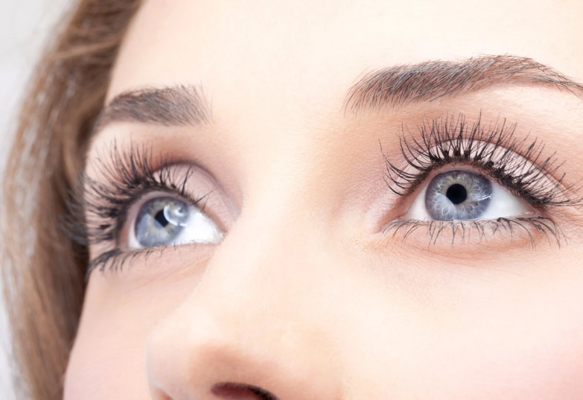
Dry Eye Diagnosis – Send Us Your Results
Learn how to get the most useful information out of your visit to the eye doctor. The results of the following tests indicate which one of the tear secretion glands is not working and what type of dry eyes you have. When performed by a physician, the results of these tests can help you select the right TheraLife® products to relieve your symptoms.Dry eyes can usually be diagnosed by the symptoms alone. Tests can determine both the quantity and the quality of the tears. A slit lamp examination can be performed to diagnose dry eyes and to document any damage to the eye.
Schirmer’s Test – Measures Tear Volume from Lacrimal Gland
This test measures the amount of moisture bathing the eye. This test is useful for determining the severity of a dry eye condition. A five-minute Schirmer’s test is performed (with or without anesthesia) using a filter paper 5 mm wide by 35 mm long. Wetting less than 5 mm with or without anesthesia is considered diagnostic for dry eyes.
If the results for the Schirmer’s test are abnormal, a Schirmer II test can be performed to measure reflex secretion. In this test, the nasal mucosa is irritated with a cotton-tipped applicator, after which tear production is measured with a filter paper. For this test, wetting less than 15 mm after five minutes is considered abnormal.
Tear Breakup Time Test – Measures Tear Viscosity & Meibomian Gland Function
A tear breakup time (TBUT) test measures the time it takes for tears to break up in the eye. The tear breakup time can be determined after placing a drop of fluorescein in the cul-de-sac of the eye.
Tear Protein Analysis
This test is not performed very often. A tear protein analysis test measures the lysozyme contained within tears. In tears, lysozyme accounts for approximately 20 to 40 percent of total protein content.
A tear lactoferrin analysis test provides good correlation with other tests.
Ask your eye doctor for the values of this test each time you visit. These values can be very useful when tracking dry eye recovery.
Best Oral Dry Eye Treatment That Works.

Add To Cart
References
1. Craig JP, Nichols KK, Akpek EK, Caffery B, Dua HS, Joo CK, et al. TFOS DEWS II definition and classification report. Ocul Surf. (2017) 15:276–83. doi: 10.1016/j.jtos.2017.05.008
2. Tchegnon E, Liao CP, Ghotbi E, Shipman T, Wang Y, McKay RM, et al. Epithelial stem cell homeostasis in Meibomian gland development, dysfunction, and dry eye disease. JCI Insight. (2021) 6:e151078. doi: 10.1172/jci.insight.151078
3. Eom Y, Lee JS, Keun Lee H, Myung Kim H, Suk Song J. Comparison of conjunctival staining between lissamine green and yellow filtered fluorescein sodium. Can J Ophthalmol. (2015) 50:273–7. doi: 10.1016/j.jcjo.2015.05.007
4. Chen M, Miki M, Lin S, Yung Choi S. Sodium Fluorescein staining of the cornea for the diagnosis of dry eye: a comparison of three eye solutions. Med Hypothesis Discov Innov Ophthalmol. (2017) 6:105–9.
5. Ding JE, Kim YH Yi SM, Graham AD Li W, Lin MC. Ocular surface cooling rate associated with tear film characteristics and the maximum interblink period. Sci Rep. (2021) 11:15030. doi: 10.1038/s41598-021-94568-9
6. Alishahi M, Kamali R. Forced diffusion of water molecules through aquaporin-5 biomembr a molecular dynamics study. Biophys Physicobiol. (2018) 15:255–62. doi: 10.2142/biophysico.15.0_255
7. van Setten GB. Osmokinetics: A new dynamic concept in dry eye disease. J Fr Ophtalmol. (2019) 42:221–5. doi: 10.1016/j.jfo.2018.11.001
8. Fagehi R, El-Hiti GA, Alqarni BM, Alanazi MA, Masmali AM, Almubrad T. Improvement in tear ferning patterns of sheep tears after addition of various electrolyte solutions. Front Med. (2021) 8:721969. doi: 10.3389/fmed.2021.721969
9. van Setten GB. Impact of attrition, intercellular shear in dry eye disease: when cells are challenged and neurons are triggered. Int J Mol Sci. (2020) 21:4333. doi: 10.3390/ijms21124333
10. Guzmán M, Miglio M, Keitelman I, Shiromizu CM, Sabbione F, Fuentes F, et al. Transient tear hyperosmolarity disrupts the neuroimmune homeostasis of the ocular surface and facilitates dry eye onset. Immunology. (2020) 161:148–61. doi: 10.1111/imm.13243
11. Hori J, Kunishige T, Nakano Y. Immune checkpoints contribute corneal immune privilege: implications for dry eye associated with checkpoint inhibitors. Int J Mol Sci. (2020) 21:3962. doi: 10.3390/ijms21113962
12. Aragona P, Giannaccare G, Mencucci R, Rubino P, Cantera E. Rolando M. Modern approach to the treatment of dry eye, a complex multifactorial disease: a PICASSO board review. Br J Ophthalmol. (2021) 105:446–53. doi: 10.1136/bjophthalmol-2019-315747
13. Chinese Branch of the Asian Dry Eye Soci Ocular Surface and Tear Film Diseases Group of Ophthalmology Committee of Cross-Straits Medicine Exchange Associate Ocular Surface and Dry Eye Group of Chinese Ophthalmologist Association. [Chinese expert consensus on dry eye: dry eye related to immunologic diseases (2021)]. Zhonghua Yan Ke Za Zhi. (2021) 57:898–907. doi: 10.3760/cma.j.cn112142-20210726-00350
14. Chinese Optometric Association of Chinese Ophthalmological Soci Optometry Group of Chinese Ophthalmologist Associate Refractive Surgery Group of Chinese Ophthalmologist Association. [Expert consensus on the diagnosis and treatment of dry eye during perioperative period of corneal refractive surgery in China (2021)]. Zhonghua Yan Ke Za Zhi. (2021) 57:644–50. doi: 10.3760/cma.j.cn112142-20210312-00124
15. Saldanha IJ, Petris R, Makara M, Channa P, Akpek EK. Impact of the COVID-19 pandemic on eye strain and dry eye symptoms. Ocul Surf. (2021) 22:38–46. doi: 10.1016/j.jtos.2021.06.004
16. Starr CE, Dana R, Pflugfelder SC, Holland EJ, Zhang S. Owen D, et al. Dry eye disease flares: a rapid evidence assessment. Ocul Surf. (2021) 22:51–9. doi: 10.1016/j.jtos.2021.07.001
17. Shen Lee B, Kabat AG, Bacharach J, Karpecki P, Luchs J. Managing dry eye disease and facilitating realistic patient expectations: a review and appraisal of current therapies. Clin Ophthalmol. (2020) 14:119–26. doi: 10.2147/OPTH.S228838
19. Schirmer O. Studien zur physiologie und pathologie der tranen-absonderung und tranenabfuhr. Graefes Arch Clin Exp Ophthalmol. (1903) 56:197–291. doi: 10.1007/BF01946264
20. Rodney WM, Louie J, Puffer JC. Schirmer’s test of lacrimation. Am Fam Physician. (1981) 24:161–4.
21. Savini G, Prabhawasat P, Kojima T, Grueterich M, Espana E, Goto E. The challenge of dry eye diagnosis. Clin Ophthalmol. (2008) 2:31–55. doi: 10.2147/OPTH.S1496
22. Shapiro A, Merin S. Schirmer test and break-up time of tear film in normal subjects. Am J Ophthalmol. (1979) 88:752–7. doi: 10.1016/0002-9394(79)90678-0
23. JordanA BaumJ. Basic tear flow. Does it exist? Ophthalmology.1980 87:920–930. doi: 10.1016/S0161-6420(80)35143-9
24. Lamberts DW, Foster CS, Perry HD. Schirmer test after topical anesthesia and the tear meniscus height in normal eyes. Arch Ophthalmol. (1979) 97:1082–5. doi: 10.1001/archopht.1979.01020010536004
25. Jones LT. The lacrimal secretory system and its treatment. Am J Ophthalmol. (1966) 62:47–60. doi: 10.1016/0002-9394(66)91676-X
27. Clinch TE, Benedetto DA, Felberg NT, Laibson PR. Schirmer’s test. A closer look. Arch Ophthalmol. (1983) 101:1383–6. doi: 10.1001/archopht.1983.01040020385009
28. Saleh TA, McDermott B, Bates AK, Ewings P. Phenol red thread test vs Schirmer’s test: a comparative study. Eye. (2006) 20:913–5. doi: 10.1038/sj.eye.6702052
29. Li N, Deng XG, He MF. Comparison of the Schirmer I test with and without topical anesthesia for diagnosing dry eye. Int J Ophthalmol. (2012) 5:478–81. doi: 10.3980/j.issn.2222-3959.2012.04.14
Learn how to get the most useful information out of your visit to the eye doctor. The results of the following diagnostic tests indicate which one of the tear secretion glands is not working and what type of dry eyes you have. When performed by a physician, the results of these tests can help you select the right TheraLife® products to relieve your symptoms.Dry eyes can usually be diagnosed by the symptoms alone. Tests can determine both the quantity and the quality of the tears. A slit lamp examination can be performed to diagnose dry eyes and to document any damage to the eye.
Schirmer’s Test – Measures Tear Volume from Lacrimal Gland
This test measures the amount of moisture bathing the eye. This test is useful for determining the severity of a dry eye condition. A five-minute Schirmer’s test is performed (with or without anesthesia) using a filter paper 5 mm wide by 35 mm long. Wetting less than 5 mm with or without anesthesia is considered diagnostic for dry eyes. If the results for the Schirmer’s test are abnormal, a Schirmer II test can be performed to measure reflex secretion. In this test, the nasal mucosa is irritated with a cotton-tipped applicator, after which tear production is measured with a filter paper. For this test, wetting less than 15 mm after five minutes is considered abnormal.
Tear Breakup Time Test – Measures Tear Viscosity & Meibomian Gland Function
A tear breakup time (TBUT) test measures the time it takes for tears to break up in the eye. The tear breakup time can be determined after placing a drop of fluorescein in the cul-de-sac of the eye.
Tear Protein Analysis
This test is not performed very often. A tear protein analysis test measures the lysozyme contained within tears. In tears, lysozyme accounts for approximately 20 to 40 percent of total protein content. A tear lactoferrin analysis test provides good correlation with other tests.